Download
1 / 12
240 likes | 599 Views
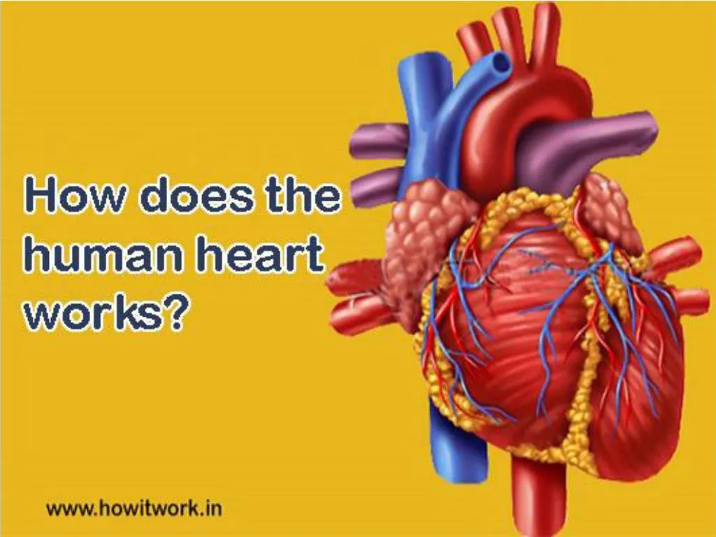
Heart is a muscular organ in humans and other animals too. The work of this heart is to pump the blood through the blood vessels of the circulatory system. This blood provides oxygen to the body and removes metabolic wastes. It is located in between the lungs and the middle compartment of the chest.

E N D
Introduction The heart is a muscular organ in humans and other animals too. The work of this heart is to pump the blood through the blood vessels of the circulatory system. This blood provides oxygen to the body and removes metabolic wastes. It is located in between the lungs an the middle compartment of the chest. www.howitwork.in
Location Theheart is located in between the lungs an the middle compartment of the chest. Heart is typically the size of a fist www.howitwork.in
Importance of Heart Heart is to pump the blood through the blood vessels of the circulatory system. This blood provides oxygen to the body and removes metabolic wastes. It is located in between the lungs an the middle compartment of the chest. www.howitwork.in
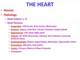
Functions of Heart Main functions of the heart can be summarised as follows : • Right - Hand Side of the Heart Right hand side of the heart receives de-oxygenated blood from the body tissues into the right atrium. • Left - Hand Side of the Heart Left-hand side of the heart receives oxygenated blood from the lungs into the left atrium. www.howitwork.in
Working In humans, mammals and birds are divided into four chambers. They are upper left and right atria and lower left and right ventricles. Commonly the right atrium and ventricles are referred together as the right heart and their left counter parts as the left heart. Fish in contrast have two chambers on atrium and a ventricle, while the reptiles have three chambers. In a active heart blood flows one way through the heart due to heart valves, which prevent the back flow. www.howitwork.in
The heart is enclosed in a protective sack, the pericardium, which also contains a small amount of fluid. The valve of the heart is made up of three layers they are epicardium, myocardium and endocardium. The heart pumps blood with a rhythm determined by a group of pace making cells in the Sino atrial node. It generate a current that causes contraction of the heart, travelling through the atrio ventricular node and along the conduction system of the heart. www.howitwork.in
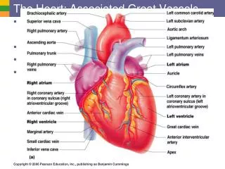
Blood flow The heart receives blood flow in oxygen from the systemic circulation, which enters the right atrium from the superior and inferior vena cavae and passes to the right ventricle. It is pumped into the pulmonary of circulation, and then through the lungs and where it receives oxygen and gives of carbon dioxide. The heart has four valves which separate its chambers. One valve lies between each atrium and ventricle, and one valve rests at the exit of each ventricle. The valves between atria and ventricles are called the atrio ventricular valves. www.howitwork.in
Between the right atrium and the right ventricle is the tricuspid valve. The tricuspid valve has three cups, which connect to chordae tendinae and three papillary muscles named the anterior, posterior, and septal muscles, after their relative positions. The mitral valve lies between the left atrium and left ventricle. It is also known as the bicuspid valve. The papillary muscles extend from the walls of the heart to valves by cartiligagenous connections called cadre tendinae. These muscles prevent the valves from falling too far back when they close. During the relaxation phase of the cardiac cycle, the papillary muscles are also relaxed and the tension on the cadre tendineae is slight. As the heart chambers contact, so do the papillary muscles. This creates tension in the cardae tendineae. www.howitwork.in
Advantages Pumps the blood Transports the blood to every part of the body. www.howitwork.in
Disadvantages Cannot be replaced Very sensitive part. www.howitwork.in