Download
1 / 20
310 likes | 1.23k Views
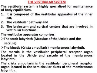
The Large Vestibular Aqueduct Syndrome in adults. an almost understimated realm Dr. Dieter Goettmann Stuttgart University Medical Centers Groningen, Nijmegen. Preface. LVAS is a syndrom Sensorineural hearing loss Sudden Fluctuating Progressive Imaging subsidary. Anatomy – Historic.

E N D
The Large Vestibular Aqueduct Syndrome in adults an almost understimated realm Dr. Dieter Goettmann Stuttgart University Medical Centers Groningen, Nijmegen
Preface • LVAS is a syndrom • Sensorineural hearing loss • Sudden • Fluctuating • Progressive • Imaging subsidary
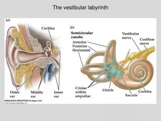
Anatomy – Historic E.Pernkopf: Atlas der topographischen Anatomie des Menschen, Bd.IV,München 1960, Tafel 162
Anatomy - Terminology • Endolymphatic sac • Extraosseus part • Intraosseus part • Commonly interpretated as the endoymphatic duct • Preductal • Endolymphatic duct • Small & short • Alsmost never dilatated
Anatomy – Terminology Intraosseus Part External Aperture of Vestibular Aqueduct Extraosseus Part of Endolymphatic Sac
Endolymph Unique extracellular fluid • High potassium: 157 mM (CSF: 3.1 mM) • Paramagnetic • Low sodium: 1.3 mMol (CSF: 149 mM) • Low calcium: 0.023 mM (Perilymph 0.6 -1.3 mM) • High electric Potential: 85 mV (CSF: 0mV) • Endocochlear potential • Low flow rate Wangemann, P. and Schacht, J. (1996) Cochlear homeostasis. In: P. Dallos, A.N. Popper and R.R. Fay (Eds) The Cochlea. Handbook of Auditory Research. Vol. 8, Springer, pp. 130-185.
1. Widened • Pre-formed • Becoming symptomatic lately
1. Widened Duct * "funnel"-shape of the extraosseus portion of the endolymphatic sac
2. Enlarging • Changes in size and/or delineation of the endolymphatic duct • Clue: Sclerosis of surrounding bone • Fuzzy, hypoattenuated border
3. Third Window • Superior Semicircular Canal Dehiscience • Fenestration or thinned bony layer of the superior semicircular canal1 • Vestibular symptoms evoked by • Sound • Pressure applied to the external auditory canal • Frequency (path., n=1000)2 • Complete defect (fenestration) 0.5 % • 4/5 superior petrosal sinus • Thinned < 0.1 mm: 1,4 % LB Minor, D Salomon, JS Zinreich, DS Zee: Sound- and/or Pressure-Induced Vertigo Due to Bone Dehiscience of The Superior Semicircular Canal. Arch Otolaryngol 1998(124):249-258 2. JP Carey, LB Minor, GT Nager: Dehiscience or Thinning of Bone Overlying Superior Semicircular Canal in a Temporal Bone Survey. Arch Otolaryngol 2000(126): 137-147
SSCD (?) • Superior petrosal sinus groove • Partial volume effect Superior Petrosal Sinus Groove
4.High Jugular Bulb Variants • Irregular shape
Jugular Bulb Diverticulum • High flow in the jugular bulb may cause excavation (wall shear stress -> remodelling) • Sometimes it reaches structures of the inner ear • Posterior semicircular canal • Endolymphatic sac Posterior SCC Endolymphatic sac
High Riding Jugular Bulb • Fuzzy margins, not to be explained by partial volume effects • Sclerosis Endolymphatic sac (intraosseus segment) Normal Margin
Symptoms of Jugular Bulb Anomalies • Sensorineural hearing loss • Conductive hearing loss • Tinnitus • Pulsatile • Often not well differentiated by the Otologist • E.g. „progressieve sensoneural hearing loss with a conductive component“
Hints • Think of it • In the field of SNHL, CT can provide more answers than MRI • Size (ax.) of the intraosseus part • Arbitrary: <= 1,5 mm, <= size f posterior SCC • Deliniation • Fuzziness • Environmental sclerotic reaction • Jugular bulb • High? High riding? • Divertikel? (coronal plane) • Arrosion of the inner ear?
Conclusion • LVAS is a syndrome with enlargement being just one feature • In adults it may have different imaging features than in children e.g. • Enlargement of the endolymphatic sac • May be progressive • Surrounding sclerotic reaction • Dehiscience of a semicircular canal • Clinical correlation w. evoked vestibular symptoms essential • Jugular bulb variants • Jugular Bulb Diverticulum • High riding Jugular Bulb • Understanding of physiology is reconsidered. As is pathophysiology.