Download
1 / 88
920 likes | 1.45k Views
The Cardiovascular System. The major organs of the cardiovascular system The heart structure and function. After today you should be able to: For more help: Chapter13 pp. 329-364. Name the organs of the cardiovascular system and discuss their functions.

E N D
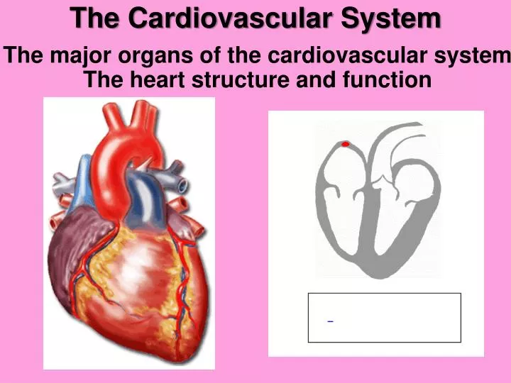
The Cardiovascular System The major organs of the cardiovascular systemThe heart structure and function
After today you should be able to: For more help: Chapter13 pp. 329-364 • Name the organs of the cardiovascular system and discuss their functions. 2. Name and describe the locations and functions of the major parts of the heart. 3. Trace the pathway of the blood through the heart and the vessels of the coronary circulation.
Major organs of the cardiovascular system • The heart • Arteries – strong elastic vessels that carry blood away from the heart. • A common misconception is that all arteries carry oxygen-rich blood.
Major organs of the cardiovascular system • Capillaries: Arteries/arterioles branch into capillaries. • They are extremely narrow, microscopic tubes with a wall that is only comprised of epithelium. • Veins- carry blood back to the atria of the heart following pathways that are almost parallel to the arteries.
Did you know… The Heart In the course of a lifetime, a human heart can beat over two billion times. • Composed of cardiac muscle tissue (myocardium) • Surrounded by a pericardium (thick membranous sack that supports and protects the heart) • Is a cone shaped, muscular organ located medially between the lungs and deep to the breastbone (sternum).
The Heart • The heart is divided into four chambers: • The LEFT and RIGHT ATRIA • The LEFT and RIGHT VENTRICLES • There are four distinct valves. • The valves actually create the beating sound of the heart.
The Heart: Right side • Takes in deoxygenated (oxygen poor blood) from the body to the heart. • Begins with the Vena Cavas.
Superior Vena Cava vein bringing de-oxygenated blood from the upper body to the heart and empties into the right atrium.
Inferior Vena Cava • vein bringing de-oxygenated blood from the lower body to the right atrium of the heart. What is meant by de-oxygenated blood? Why is the blood de-oxygenated?
Why is the blood de-oxygenated? • Inside the cell, the mitochondria uses the O2 for cellular respiration. • During cell respiration, the O2 binds to a carbon, and is now CO2. • CO2 diffuses into the blood stream and flows back to the heart. • De-oxygenated blood = oxygen poor or has less O2 than CO2 • The oxygen that diffused from the alveoli into the blood gets delivered to the cells of the body.
Right Atrium • receives de-oxygenated blood from the body through the superior vena cava and inferior vena cava .
Tricuspid Valve • separates the right atrium from the right ventricle. • It opens to allow the de-oxygenated blood from the right atrium to flow into the right ventricle and prevents blood from returning to the right atrium.
Right Ventricle • receives de-oxygenated blood from the right atrium and pushes it next through the pulmonary valve.
Pulmonary Valve • separates the right ventricle from the pulmonary artery. • Allows blood to flow from the Right ventricle to the pulmonary arteries.
Pulmonary Artery • is the vessel transporting de-oxygenated blood from the right ventricle to the lungs.
Summarize what we know so far: ANSWER THE FOLLOWING QUESTIONS: • Where does deoxygenated blood originate from? • Where in the heart does the deoxygenated blood enter first? • Where does the deoxygenated blood go next? • What two valves are on the right side of the heart? What are the roles of these 2 valves? • Where does blood exit and go to from the right side of the heart? • Is it de-oxygenated (oxygen poor) or oxygenated (oxygen rich)?
STOP! • Label the right side of the heart only on the heart diagram.
The Heart: Left side • Brings oxygenated (oxygen rich blood) from the lungs to the heart. • Begins with the Pulmonary Vein.
Pulmonary Vein • is the vessel transporting oxygen-rich bloodfrom the lungs to the left atrium.
Left Atrium • receives oxygenated blood from the lungs from the pulmonary vein
Bicuspid Value • separates the left atrium from the left ventricle. • It opens to allow the oxygenated blood to flow into the left ventricle and prevents it from flowing back.
Left Ventricle • receives oxygenated blood as the left atrium contracts and the bicuspid valve opens.
Aortic Valve • separates the left ventricle from the aorta. • As the ventricles contract, it opens to allow the oxygenated blood collected in the left ventricle to flow throughout the body and prevents it from going back to the heart.
Aorta • is the largest single blood vessel in the body. • This vessel carries oxygen-rich blood from the left ventricle to the various parts of the body.
Summarize what we know so far: ANSWER THE FOLLOWING QUESTIONS: • Where does oxygenated blood originate from? • Where in the heart does the oxygenated blood enter first? • Where does the oxygenated blood go next? • What two valves are on the left side of the heart? What are the roles of these 2 valves? • Where does blood exit and go to from the left side of the heart? • Is it de-oxygenated (oxygen poor) or oxygenated (oxygen rich)?
Papillary Muscles • Papillary muscles: attach to the lower portion of the interior wall of the ventricles. • They connect to the chordae tendineae on the valves, • The contraction of the papillary muscles opens the valves. When the papillary muscles relax, the valves close.
Chordae Tendineae • Chordae tendineae are tendons linking the papillary muscles to the tricuspid valve in the right ventricle and the mitral valve in the left ventricle. • The chordae tendineae are string-like in appearance and are sometimes referred to as "heart strings."
Ventricular Septum • Ventricular Septum: wall separating the lower chambers (the ventricles) of the heart from one another.
Heart Activities: • Finish cardiovascular diagram • Complete organ chart. • Create Heart Foldable • Vocab Index Card Blood flow order activity
Electrical Conduction Pathway: Be Still My Beating Heart
The Lub-Dub… • Heartbeat is the sound you hear when the valves of the heart close. • Each heartbeat is called a cardiac cycle. • Controlled by the electrical conduction pathway • First the Atria contract at the same time sending the blood to the ventricles. • Then the ventricles contract at the same time sending blood to the pulmonary artery or the aorta. • http://www.youtube.com/watch?v=v3b-YhZmQu8
The Lub-Dub… • Diastole is the relaxing phase of the heart – when the chambers are resting. • Systole is the working phase of the heart – when the chambers contract.
The Lub-Dub… • Dup – the relaxation of the ventricles causes blood to flow backward momentarily and the pulmonary and aortic valves close. • Lub – is the sound you hear when the blood pressure increases in the ventricles forcing the tricuspid and bicuspid valves to slam shut but causing the pulmonary and aortic valves to open.
Murmurs… • Swishing sound after the lub • Leaky valves allows blood to flow back into the atrias. • The two types of heart murmurs are innocent (harmless) and abnormal.
Murmurs… • Innocent murmurs are simply sounds made by blood flowing through the heart's chambers and valves, or through blood vessels near the heart. • Congenital heart defects or acquired heart valve disease often are the cause of abnormal heart murmurs.
Electrical Conduction Pathway: Be Still My Beating Heart
serves as the natural pacemaker for the heart. Nestled in the upper area of the right atrium, it sends the electrical impulse that triggers each heartbeat. The impulse spreads through the atria, coordinated wave-like manner. Sinoatrial Node (often called the SA node or sinus node)
The impulse that originates from the SA node strikes AV node situated in the lower portion of the right atrium. The AV node in turn sends an impulse through the nerve network to the ventricles to contract. Atrioventricular node (or AV node)
electrical network serving the upper ventricles These nerve fibers send impulses that cause the cardiac muscle tissue to contract. Right and Left Bundle Branches.
electrical network serving the lower ventricles These nerve fibers send impulses that cause the cardiac muscle tissue to contract. Purkinje Fibers
Electrical Conduction Pathway: • The SA Node to the AV Node to the left and right Bundle Branches - to the Purkinje Fibers = THE HEART BEAT and CONTRACTIONS
BLOOD PRESSURE The force blood exerts again the inner walls of the vessels
Arterial Blood Pressure • Rises and falls in a pattern corresponding to the phases of the cardiac cycle. • Contracting ventricles (ventricular systole) squeeze blood out and into the arteries – increases pressure in these vessels • This is called systolic pressure – the maximum pressure during contraction. • This value is usually around 120mmHg
Arterial Blood Pressure • When ventricles relax (ventricular diastole) , arterial pressure drops • The lowest pressure that remains is called diastolic pressure. • This value is usually around 80mmHg