Download
1 / 26
260 likes | 270 Views
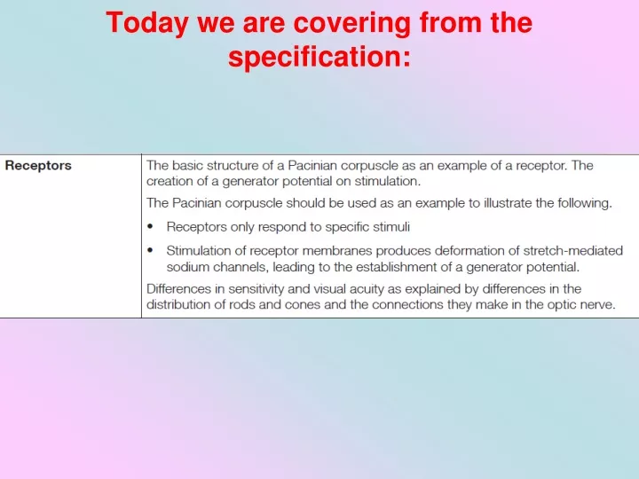
Learn about the sensory nervous system and how receptors detect changes in the body's environment. Explore different types of receptors and their functions, such as mechanoreceptors and photoreceptors.

E N D
The sensory nervous system Receptors are specialized cells that can detect changes in the body’s internal and external environment. Most receptor cells are only sensitive to one type of stimulus. Receptors convert the energy of the stimulus into the start of a nerve impulse known as a generator potential. For example, mechanoreceptors detect changes in mechanical energy, such as pressure. mechanoreceptor in the skin
Read the article ‘How are you feeling?’. • Answer these questions: • What is the main difference between a Meissner’s corpuscle and a Merkel’s disc? • What is a ‘two point discrimination test’? • Explain how a mechanoreceptor converts mechanical energy into electrical energy. • Explain how a Pacinian corpuscle can produce a generator potential. • List some examples of the uses of haptic technology.
Receptors - Pressure A Pacinian corpuscle Yteach resource
Pacinian corpuscle Layers of connective tissue separated by a gel Blood capillary
Stretch-mediated sodium channels (Permeability change when shape changes) Resting Potential: Positive outside – negative inside Pressure: Distorts & opens Na+ channels Generator potential: Inflow of Na+ depolarises membrane
How do rod cells produce impulses? Rod cells allow vision in dim light due to the presence of a pigment called rhodopsin, which is found in membrane-bound vesicles. vesicles containing rhodopsin When rhodopsinabsorbs light it splits into its constituent parts, opsin and retinal. This is called bleaching. Low intensity light is sufficient to cause this breakdown. The presence of opsin causes a change in the permeability of the rod cell to sodium, which initiates a generator potential. Rhodopsin can reform in the absence of further light stimulation.
How do cone cells produce impulses? Cone cells aresensitive to high light intensities due to the presence of the pigment iodopsin. vesicles containing iodopsin In bright light, iodopsin is broken down into its constituent parts, generating an action potential in the ganglion cell. There are three different types of cone cell, each containing a different form of iodopsin. Each form of iodopsin absorbs a different wavelength of light – green, blue or red. The colour seen depends on the relative degree of stimulation of the three different types of cone cell.
Photoreceptors These are found in the retina. There are two types Rods and Cones and they are arranged as shown: Outer ! Inner Light Pigmented Layer Ganglion Cells Rod To Optic Nerve Bipolar Neurones Cone
Light receptors Both rod and cone cells act as transducers by converting light energy into the electrical energy of a nerve impulse.
Rod cells Cannot distinguish different wavelengths of light and therefore produce images only in black and white. Rod cells are more numerous than cones.
Rod cells Many rod cells share a single sensory neurone. Rod cells can therefore respond to light of very low intensity. This is because a certain threshold value has to be exceeded before a generator potential is created in the bipolar cells to which they are attached.
Rod cells A number of rod cells are attached to a single bipolar cell (= retinal convergence), there is a much greater chance that the threshold value will be exceeded than if only a single rod cell were attached to each bipolar cell. As a result, rod cells allow us to see in low light intensity (i.e. at night), although only in black and white.
Changes in the electrical potential of a receptor when stimulated by three separate stimuli. Only the third stimulus produces a generator potential high enough to trigger a nerve impulse.
A rod cell LIGHT opsin Rhodopsin (pigment in rod cells broken down) Signal from Bipolar cell
A rod cell As many rod cells are joined to the same bipolar cells, only a single impulse will be stimulated. This means that they cannot distinguish between the separate sources of light that stimulated them. 2 dots close together will appear as a single blob. Rod cells therefore have low visual acuity.
Cone cells Cone cells are of three different types, each responding to a different wavelength of light. Depending on the proportion of each type that is stimulated, we can perceive images in full colour. Each cone cell usually has its own bipolar cell connected to a sensory neurone. This means that often the generator potential is not exceeded. As a result, cone cells only respond to high light intensity and not to low light intensity.
Cone cells Cone cells contain a different pigment to rod cells (iodopsin). This requires a higher light intensity to be broken down and create a generator potential. As cone cells are attached to their own bipolar cell, if 2 adjacent cells are stimulated, the brain receives 2 separate impulses. Cone cells give very accurate vision, they have good visual acuity.
Cone cells Light is focussed by the lens on a point known as the fovea. The fovea therefore receives the highest intensity of light. Therefore cone cells, but not rod cells, are found at the fovea. The concentration of cone cells diminishes further away from the fovea. At the peripheries of the retina, where light intensity is at its lowest, only the rod cells are found.
Distribution of Rods and Cones • Fovea – area of retina where light is focused – highest concentration of cones – few elsewhere. • Rods most concentrated either side of fovea but still many elsewhere. • No rods or cones at blind spot – this is where the optic nerve exits the eye.
Colour Blindness • If you have normal vision you will see a figure seven in reddish brown dots. • People with red-green colour blindness will not see the 7, why? • These people lack red sensitive cones, but the green stimulated cones are stimulated by the red light, so all dots appear green
Further Questions • Explain why brightly coloured objects often appear grey in dim light. • At night, it is often easier to see a star in the sky by looking slightly to the side of it rather than directly at it. Suggest why this is so.