Download

1 / 17
190 likes | 405 Views
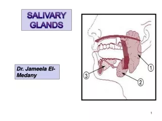
The parotid, which is the larges t gland, is located anterior to the external acoustic meatus and mastoid process , inferior to the zygomatic arch , lateral and posterior to the ramus of the mandible, and on the surface of the masseter muscle .

E N D
The parotid, which is the larges t gland, is located anterior to the external acoustic meatus and mastoid process, inferior to the zygomatic arch, lateral and posterior to the ramus of the mandible, and on the surface of the masseter muscle. • Anatomically, the Parotid gland is closely associated with the facial nerve, external carotid artery, superficial temporal and maxillary veins and numerous cervical lymph nodes.
The parotid (Stensen's ) duct extends from the lateral surface of the gland, anteriorly, across the masseter muscle and the buccal fat pad. • At the anterior border of the masseter, it bends medially at a sharp angle, piercing the fat pad and buccinator muscle to open into the oral cavity in a papilla opposite the crown of the second maxillary molar tooth. The epithelium of the duct becomes continuous with the mucous membrane of the mouth.
The submandibular gland is located medial to, and under partial cover of the mandible. It is closely associated with the mylohyoid and medial pterygoid muscles, submandibular lymph nodes and facial arteries and veins. • The submandibular (Wharton's) duct extends anteriorly in the floor of the mouth to open into the oral cavity at the sublingual papilla at the side of the frenulum of the tongue.
The sublingual is the smallest of the major salivary glands and is Iocated beneath the mucous membrane of the floor of the mouth. • Although the parotid and submandibular glands are encased in an extensive connective tissue capsule, the sublingual gland lacks a distinct capsule. • compared with the parotid and submandibular the sublingual consists of large portion and collection of small glands rather than a single, clearly delineated.
The main excretory duct of sublingual gland (Bartholin's) may join the submandibular duct or open into the oral cavity with a separate sublingual papilla. • Numerous smaller sublingual ducts (ducts of Rivinus) may join the submandibular duct or open separately into the floor of the mouth.
Histology of the Salivary Glands • The salivary glands are composed of large numbers of secretory acini, which may be tubular or globular in shape. • Each acinus drains into a duct. These microscopic ducts coalesce to form lobular ducts. • Each lobule has its own duct and these then merge to form the main ducts. The individual lobes and lobules are separated by dense connective tissue, which is continuous with the gland capsule. • The ducts, blood vessels, lymphatics, and nerves run through and are supported by this connective tissue.
The acini are the primary secretory organs but the saliva is modified as it passes through the intercalated, striated, and excretory ducts before being discharged into the mouth and oropharynx (Figure1.14). • The lobules also contain significant amounts of adipose tissue particularly in the parotid gland. • The proportion of adipose tissue relative to excretory acinar cells increases with age.
In the human parotid, the excretory acini are almost entirely serous. In the submandibular gland, again, the secretory units are mostly serous but there are additional mucous tubules and acini. • In some areas the mucinous acini have crescentic “caps” of serous cells called serous demilunes. In the sublingual gland the acini are almost entirely mucinous, although there are occasional serous acini or demilunes.
The serous cells contain numerous proteinaceous secretory (zymogen) granules. These granules contain high levels of amylase. In addition, the secretory cells produce kallikrein, lactoferrin, and lysozyme. In mucous cells, the cytoplasm is packed with large pale secretory droplets.
Initially the secretory acini drain into intercalated ducts. These function mainly to conduct the saliva but they may also modify the electrolyte content and secrete immunoglobulin A. The intercalated ducts drain into striated ducts, which coalesce into intralobular and extralobular collecting ducts. • The intercalated duct cells are very active metabolically and they transport potassium and bicarbonate into saliva. They reabsorb sodium and chloride ions so that the resulting saliva is hypotonic. They also secrete immunoglobulin A, lysozyme, and kallikrein. The immunoglobulin is produced by plasma cells adjacent to the striated duct cells and it is then transported through the epithelial lining into the saliva. The main collecting ducts are simple conduits for saliva and do not modify the composition of the saliva.
Myoepithelial cells are contractile cells closely related to the secretory acini and also much of the duct system. The myoepithelial cells lie between the basal lamina and the epithelial cells. • Numerous cytoplasmic processes arise from them and surround the serous acini as basket cells. Those associated with the duct cells are more fusiform and are aligned along the length of the ducts. The cytoplasm of the myoepithelial cells contains actin myofilaments, which contract as a result of both parasympathetic and sympathetic activity. • Thus the myoepithelial cells “squeeze” the saliva out of the secretory acini and ducts and add to the salivary secretory pressure.