Download
1 / 37
540 likes | 2.12k Views
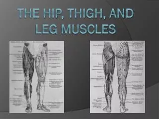
Hip Muscles. Mazyad Alotaibi. Testing Muscles of the Lower Extremity. Hip Flexion. Hip flexion, abduction and external (lateral) rotation with knee flexion. Hip Extension. Hip abduction. Hip Abduction from flexed position Hip Adduction Hip External Rotation Hip Internal rotation.

E N D
Hip Muscles MazyadAlotaibi
Testing Muscles of the Lower Extremity • Hip Flexion. • Hip flexion, abduction and external (lateral) rotation with knee flexion. • Hip Extension. • Hip abduction. • Hip Abduction from flexed position • Hip Adduction • Hip External Rotation • Hip Internal rotation
Hip Flexion 1- ANATOMY: Agonist / Prim mover : Psoasmajor and iliacus Origin: Psoas major: transverse processes of L1-L5 and the vertebral bodies of T12-L5 Iliacus: anterior 2/3 of iliac fossa Insertion: Psoas major: lesser trochanter of the femur Iliacus: lesser trochanter of the femur Nerve Supply: Psoas major: lumbar plexus , nerve root from L2-L4 Iliacus: lumbar plexus, Femoral nerve L2-L3 Action: powerful hip flexion Synergist / Accessory Muscles: Rectus Femoris (RF), Sartorius, Tensor fasciae latae (TFL). 2- Range of motion: 0 to 1200
3- Stabilization: 1. contraction of anterior abdominal muscles to fix lumbar spine and pelvis. 2. weight of trunk. 4- Effect of weakness and contracture: 1- Difficulty in: climbing stair, walking up or down the incline, getting up from a reclined position. 2- In marked weakness: walking is difficult because the leg must brought forward by pelvic motion. 3- Effect of contracture: Bilateral– Increased lumbar lordosis. Unilateral–hip abduction combined with external rotation. 5- Factor Limiting of motion: - With knee flexed, contact of thigh on abdomen. - With knee extended, tension of Hamstring Muscles. 6- Substitution: *Sartorius: external rotation and abduction of the hip *Tensor fasciae latae: internal rotation and abduction of the hip.
7-Procedures: a- Position of Patient: b- Position of Therapist : inner hand, Outer hand, Direction of Resistance c- Test d- Instruction to patient The patient is short sitting with thighs fully supported and legs hanging over the edge. The therapist stands next to the test leg. The therapist places one hand on the distal thigh and proximal knee, and applies resistance in a downward direction as the patient actively flexes at the hip
Hip flexion, abduction, and external rotation with knee flexion 1- ANATOMY: - Agonist / Prim mover :Sartorius Origin: anterior superior iliac spine (ASIS) Insertion: upper medial surface of body of tibia Action: - flexes hip and knee - With flexed hip, laterally rotates the thigh Nerve supply: branches of femoral nerve, L2-L3 Synergist / Accessory Muscles: hip and knee flexors. hip external rotators, and hip abductor. 3. Nerve supply: Femoral n.(L2-L3) 2- Range of motion: NO specific ROM because of two-joint muscle.
3- Fixation: a. Contraction of abdominal muscles to fix pelvis. b. Weight of trunk. 4-Effect of weakness and contracture: effect of weakness: loss of antro- medial instability of the knee joint. effect of contracture: flexion, abduction and lat. Rot. Deformity of the hip with knee flexion. 5- Factor Limiting of motion: Non, because incomplete range of motion. 6- Substitution: Iliopsoas or the Rectus Femoris results in pure hip flexion without abduction and external rotation.
7- Procedures: a- Position of Patient: b- Position of Therapist : inner hand, Outer hand, Direction of Resistance c- Test d- Instruction to patient. The patient is short sitting with thighs supported on table and legs hanging over side. The therapist stands lateral to the test leg while placing one hand on the lateral side of the knee and using the other hand to grasp the medial anterior surface of the distal leg. Hand at knee gives downward and inward resistance. Hand at ankle gives upward and outward resistance. Patient flexes, abducts, and externally rotates the hip and flexes the knee.
Hip Extension • ANATOMY: Prim mover / agonist: Gluteus maximus and Hamstring Origin of Gluteus maximus • outer rim of ilium (medial aspect) • dorsal surface of sacrum and coccyx • sacrotuberous ligament Insertion of Gluteus maximus : • Illiotibial tract of fascia lata(primary insertion) • gluteal tuberosity of femur Action of Gluteus maximus : • powerful extensor of hip • laterally rotates thigh • upper fibers aid in abduction of thigh • fibers of IT band stabilize a fully extended knee Nerve supply of Gluteus maximus : inferior gluteal nerve, L5,S1,S2 Synergist / Accessory Muscles: Adductor magnus (inferior part), Gluteus medius (post part), 2- Range of motion: 0 to 200 degrees (hyper) 0 to 50
3- Fixation a. Contraction of iliocostalislumborum and quadratuslumborum muscle. b. Weight of trunk. 4- Effect of weakness and contracture: 1- Effect of weak. Bilaterally makes walking difficult., difficult in raising the trunk from foreword-bent position. 2- Patient must push themselves to an upright position by using their arms during walk. 3- Effect of contracture: walking with Hyper extension deformity. 5-Factor Limiting of motion: a. Tension of iliofemoral ligament. b. Tension of hip flexor muscles. 6-Substitution: by extending lumbar spine. Therapist must support the pelvis.
7- Procedures: a- Position of Patient: b- Position of Therapist : inner hand, Outer hand, Direction of Resistance c- Test d- Instruction to patient The patient lies prone on the table. The therapist stands on the side of the test leg, at pelvis level. One hand stabilizes the pelvis, and the other hand is placed on the distal calf. The hand on the distal calf applies resistance in a downward direction ad the patient actively extends at the hip.
Hip abduction 1- ANATOMY: Prim mover/ agonist: ( Gluteus medius and Gluteus minimus) Gluteus medius: Origin: outer aspect of ilium (between iliac crest and anterior and posterior gluteal lines) • upper fascia (AKA gluteal aponeurosis) Insertion: lateral aspect of greater trochanter of femur Action: - anterior and lateral fibers abduct and medially rotate the thigh • posterior fibers may laterally rotate thigh • stabilizes the pelvis and prevents free limb from sagging during gait Nerve: superior gluteal nerve, L4,5,S1 Gluteus minimusOrigin: outer aspect of ilium (between anterior and inferior gluteal lines) Insertion: • greater trochanter (anterior to medius) • articular capsule of hip joint Action: - abduct and medially rotate the thigh • stabilizes the pelvis and prevents free limb from sagging during gait Nerve: superior gluteal nerve, L4,5,S1
Hip abduction Synergist / Accessory Muscles: Upper fiber of Gluteus maximus, Sartorius, TFL. 2- Range of motion: 0 to 45 degrees 3- Fixation: a. Contraction of lateral abdominal muscles and latissimusdorsi. b. Weight of trunk. 4- Effect of weakness and contracture: Effect of weakness: unilateral: positive (trendlingburgh test) Bilateral: waddling gate Effect of contracture: positive Ober’s test 5- Factor Limiting of motion: • Tension of distal band of iliofemoral ligament and pubocapsular ligament. • Tension of hip adductor muscle. 6- Substitution: - Patient may “hike hip” by approximating pelvis to thorax using lateral trunk muscles. - hip external rotation with flexion. - TFL substitution
7- Procedures: a- Position of Patient: b- Position of Therapist : inner hand, Outer hand, Direction of Resistance c- Test d- Instruction to patient. The patient is side lying with test leg uppermost. The therapist stands behind the patient and stabilizes with one hand at the hip. This hand is proximal to the greater trochanter. The other hand applies resistance across the lateral surface of the knee. Patient abducts hip against downward resistance.
Hip Abduction from flexed position 1- ANATOMY: Prim mover /agonist ( Tensor Fascia Latae): Origin: - anterior aspect of iliac crest - anterior superior iliac spine (ASIS) Insertion: anterior aspect of IT band, below greater trochanter Action: - hip flexion • medially rotate & abduct a flexed thigh • tenses IT tract to support femur on the tibia during standing Nerve: superior gluteal nerve, L4,L5,S1 Synergist / Accessory Gluteus medius, and Gluteus minimus
2- Range of motion: No specific Rom, because of two-joint muscle. 3-Fixation: 1. Contraction of lateral abdominal muscles and latissimusdorsi 2. weight of trunk. 4- Effect of weakness and contracture: Effect of weakness : pt walks with Leg with tendency to rotate hip laterally Effect of shortness: Bilaterally– results in anterior pelvic tilt and sometimes bilateral knock-knees, Unilateral– results in lateral pelvic tilt. Effect of contracture: hip flexion, and knock knees. 5-Factor Limiting of motion: - Non, ROM incomplete 6-Substitution: by Hip lateral rotator muscles.
7- Procedures: a- Position of Patient: b- Position of Therapist : inner hand, Outer hand, Direction of Resistance c- Test d- Instruction to patient. The patient is side lying with test leg uppermost, and hip flexed to 45 degrees. The therapist stands behind the patient and stabilizes with one hand at the hip. This hand is proximal to the greater trochanter. The other hand applies resistance across the lateral surface of the knee. Patient abducts hip against downward resistance.
Hip Adduction 1- ANATOMY: Prim mover /agonist (Adductors magnus, Adductors Brevis, Adductors Longus,Pectineus and Gracilis) OriginInsertion Adductors magnusIschialtuberosity (inf-lat) Femur (lineaaspera) Adductors Brevis Pubis Femur (lineaaspera) Adductors Longus Pubis Femur (lineaaspera) Pectineus Pubis Femur (lineaaspera) Gracilis Pubis Femur (lineaaspera) Action: Hip Adduction Nerve supply: All Adductors are supply by Obturator nerve (L2,3,4) Pectineus is supplied by Femoral n.(L2-L3) Synergist / Accessory Muscles: Obturator externus, Gluteus maximus.
2- Range of motion: 0 to 15- 20 3- Fixation: by Weight of trunk 4- Effect of weakness and contracture: - Effect of weakness : patient unable to adduct the leg during walking. - Effect of shortness: patient walks with adducted legs. - Effect of contracture: unable to abduct leg during gate cycle. 5- Factor Limiting of motion: 1. Contact with opposite limb. 2. When hip is flexed, tension of ischiofemoral ligament 6- Substitution: by 1. hip flexor muscles. 2. Hamstring muscle.
7- Procedures: a- Position of Patient: b- Position of Therapist : inner hand, Outer hand, Direction of Resistance c- Test d- Instruction to patient. The patient is side lying with the test leg lowermost and resting on the table. The uppermost leg is abducted to 25 degrees and supported by the examiner. The therapist stands behind the patient at the knee level. The resistance hand is placed on the distal medial femur of the test leg. Resistance is applied in a downward motion while the patient actively adducts.
Hip External Rotation 1- ANATOMY: Prim mover /agonist (Obturatorsinternus and externus, Gemellae superior and inferior, Piriformis, QuadratusFemoris, Gluteus maximus “posterior” Origin Insertion ObturatorsinternusIschium and Pubis Femur (trochantericfossa ) ObturatorsexternusIschium and Pubis Femur (greater trochanter) Gemellae Superior Ischium Femur (greater trochanter) Gemellae Inferior Ischialtuberosity Femur (greater trochanter) Piriformis Sacrum Femur (greater trochanter) QuadratusFemorisIschialtuberosity Femur Gluteus Maximus Ilium , sacrum Femur (gluteal tuberosity) Nerve supply: Obturatorsinternus: Nerve to Obturatorsinternus (L5-S1) Obturatorsexternus: Nerve to Obturatorsexternus (L3-L4) Gemellae Superior: Nerve to Gemellae Superior (L5-S1) Gemellae Inferior: Nerve to Gemellae Inferior (L5-S1) Piriformis: Nerve to Piriformis (S1-S2) QuadratusFemoris: Nerve to QuadratusFemoris (L5-S1) Gluteus Maximus: Inferior gluteal n.(L5-S2) Action: Hip lateral rotation Synergist / Accessory Muscles: Sartorius, Biceps femoris, Adductors magnus and longus
2- Range of motion: 0 to 45 3- Fixation: by Weight of trunk 4- Effect of weakness and contracture: - Effect of weak : result in medial rot. accompaied by foot pronation with knock-knees. - Effect of contracture: result in abduction position with limited medial rot. Of the hip accompained by outward position of the toes in standing position 5- Factor Limiting of motion: 1. Tension of lateral band of iliofemoral ligament. 2. Tension of hip medial rotator muscles. 6- Substitution: - Sartorius ( Hip flex,abd, and ext rot.)
7- Procedures: a- Position of Patient: b- Position of Therapist : inner hand, Outer hand, Direction of Resistance c- Test d- Instruction to patient. The patient is short sitting. The therapist sits on a stool or kneels beside patient. The therapist places one hand at the lateral aspect of the distal thigh and applies resistance in a medial direction. The other hand grasps the medial ankle just above the malleolus, and applies resistance in a lateral direction. The patient is actively externally rotating at the hip.
Hip Internal Rotation 1- ANATOMY: Prim mover /agonist ( Gleteiminimus and medius, Tensor fascia latae): Origin Insertion Gluteus MinimusIlium (outer surface) Femur (greater trochanter) Action: Hip medial rotation Nerve supply: Gluteus Minimus: Superior gluteal n.(L4-S1) Action : Hip medial rotation Synergist / Accessory Muscles: Tensor fascia latae, Gluteus minimus and medius.
2- Range of motion: 0 to 45 3- Fixation: - Weight of trunk 4- Effect of weakness and contracture: - walking with lat. Rot. 5- Factor Limiting of motion: 1. when hip is extended, tension of iliofemoral Ligament. 2. when hip is flexed, tension of ischiocapsular ligament. 3. tension of hip lateral rotator muscles. 6- Substitution: by lifting the pelvis on the tested side.
7- Procedures: a- Position of Patient: b- Position of Therapist : inner hand, Outer hand, Direction of Resistance c- Test d- Instruction to patient. The patient is short sitting. The therapist sits on a stool or kneels beside patient. The therapist places one hand at the medial aspect of the distal thigh and applies resistance in a lateral direction. The other hand grasps the lateral ankle just above the malleolus, and applies resistance in a medial direction. The patient is actively internally rotating at the hip.
TRENDELENBURG SIGN • Procedure: subject assumes unilateral stance without upper extremity assistance. Examiner observes patient from behind. • Interpretation: • Normal: Hip on opposite side should rise slightly • Abnormal • Dropping of pelvis on the opposite side • Shifting center of gravity over stance leg *These findings indicate abductor weakness of stance leg
FLEXIBILITY • Thomas Test: • Procedure: Patient in supine, both knees brought to chest. Patient holds unaffected leg, keeping their back flat against the table. The tests leg is allowed to drop into extension. Next the knee is allowed to drop into flexion • Interpretation: • Hip should extend to 0 degrees; if this is not achieved, tightness of one-joint hip flexors is indicated • If able to achieve full hip extension, but note 80 degrees of knee flexion, then tightness of the two joint hip flexors (rectus femoris) is indicated • Abduction of the hip and/or external rotation of the tibia indicate ITB tightness
FLEXIBILITY • Ober’s Test: • Procedure: Patient in side-lying with test side up. The knee may extended or flexed to 90 or 30 degrees. The hip is maintained in slight extension. The test leg is abducted, then allowed to lower toward the table with the pelvis stabilized • Interpretation: • Normal: able to abduct parallel to the examining surface • Inability to adduct to parallel indicates tightness of the ITB
FLEXIBILITY • Hamstring Flexibility 1. Passive Straight Leg Raise • Normal: should achieve at least 80 degrees of hip flexion • Reproduction at 45 degrees or less may indicate lumbar radiculopathy 2. Popliteal Angle • Patient is supine with test leg’s hip flexed to 90 degrees • The knee is passively extended • Interpretation • Normal: Angle of flexion should be 15 to 20 degrees or less • Abnormal: If angle of flexion is greater than 15 to 20 degrees, this is indicative of hamstring tightness
ELY’S TEST • Procedure: Patient in prone. The knee of tested leg is flexed by the examiner • Interpretation: • Normal: Able to fully flex the knee without creating hip flexion • Abnormal: Flexion of the hip prior to full knee flexion indicates Rectus Femoris tightness