Download
1 / 14
140 likes | 145 Views
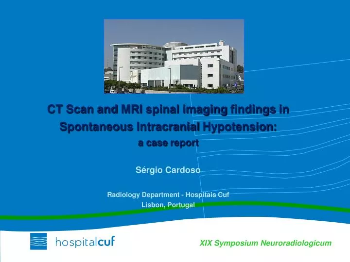
CT Scan and MRI spinal imaging findings in Spontaneous Intracranial Hypotension : a case report Sérgio Cardoso Radiology Department - Hospitais Cuf Lisbon , Portugal.

E N D
CT Scanand MRI spinalimagingfindingsinSpontaneousIntracranialHypotension: a case reportSérgio CardosoRadiologyDepartment - Hospitais CufLisbon, Portugal
Syndromeoflowcerebral spinalfluid (CSF) volume, secondary to CSF leakeageintotheepiduralspace (spinal meninges structuralweakness) No previoushistoryofsurgeryor lombar puncture; Presence of trivial traumatic events in 1/3 of patients No epidemiological data available ( annualincidence5/100.000 ) Affectsyoungandmiddle age individuals; female/male ratio 2:1 Variableclinicalpresentation ; postural headacheisthemostcommonmanifestation Delayindiagnosisisveryfrequent SpontaneousIntracranialHypotension(SIH)
34 yofemale Previouslyhealthy, with no relevantissuesinherpastmedicalhistory Suddenlyonsetofpermanentsevere, cervical painoneweekbeforefirstobservation, with no relieforexacerbationfeatures No othersymptoms, as headache, fever, systemic Inphysicalexaminationtheonlyfindingwassome degreeofneckstiffness, withotherwise normal physicalandneurologicalexamination Laboratorial workoutshowedno significantabnormalities (normal CRP, normal bloodcount, normal renal andhepaticfunction) Case Report
Cervical Spine CT Scan (03/06/2010) Antero-lateral hyperdense epidural collection
Brain MRI (03/06/2010) No abnormalfindings
Spine MRI (03/06/2010) EPIDURAL HEMATOMA ? SPINAL AVM ? Spine MRI - spinalepiduralcollection - dilatedepiduralveins - enlargementofepiduralvenousplexus - duralenhancement
SpinalAngiography • No spine AVM • No signsof medular compression • No clinicaldeterioration →conservativeattitude
Spine MRI (30/06/2010) • TypicalneuroimagingfeaturesofSIH • - epiduralfluidcollections • duralsaccollapse-festoonedappearence • duralenhancement • epiduralvenousplexusdilatation- dilatedepiduralveins • -C1\C2 sign (absent)
Dorsal and Lombar Spine MR (30/06/2010) • - collapseofduralsac • dilatationofepiduralvenousplexus
BrainMRI (30/06/2010) • Typicalbrainneuroimagingfeaturesof SIH • Subduralfluidcollections • Enhancementofpachymeninges • Engorgemntofvenousstructures • Pituitaryhyperemia • Saggingofthebrain • → SEEPS
CT Myelography(22/07/2010) Persistentsymptomswithconservativetherapy (bedrestandhydratation) → CT myelography Thedifferentialopacificationofthecalsacandthe ventral fluidcollection→signofcommunication Theexactlocationofthecommunicationcouldnotbedetermined
EVOLUTION • Conservativemeasuresuneffective (bedrest, oral hydration, caffeine intake) • Epiduralblood patch (27/07/2010) → clinicalimprovement • SpineandBrain MR (24/08/2010) • 03/06/2010 • 24/08/2010 • 30/06/2010 • 24/08/2010 24/08/2010
TakeHomeMessages • Spontaneousintracranialhypotension (SIH) isnotrare, butisstillunderdiagnosed • Clinicalandimagingfindings are widelyvariable • CT Myelographyisthestudyofchoice to identifythe CSF leak, butisnotalwaysnecessary to makethediagnosis • The role ofspinalimagingfindingsin SIH isnotwellestablished, butawareness to abnormalitiesatthislevelcanlead to anearlierdiagnosis