Download
1 / 16
160 likes | 172 Views
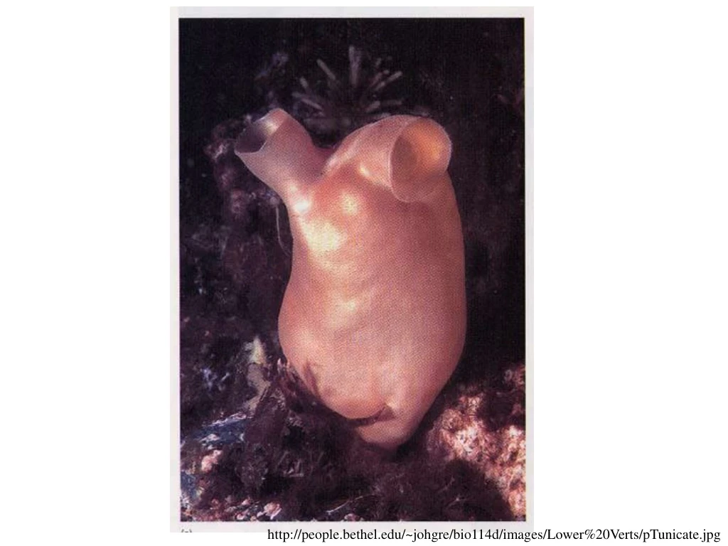
http://people.bethel.edu/~johgre/bio114d/images/Lower%20Verts/pTunicate.jpg. Figure 5.34 Bilateral symmetry in the egg of the ascidian tunicate Styela partita. Figure 5.35 Cytoplasmic rearrangement in the fertilized egg of Styela partita.

E N D
http://people.bethel.edu/~johgre/bio114d/images/Lower%20Verts/pTunicate.jpghttp://people.bethel.edu/~johgre/bio114d/images/Lower%20Verts/pTunicate.jpg
Figure 5.34 Bilateral symmetry in the egg of the ascidian tunicate Styela partita
Figure 5.35 Cytoplasmic rearrangement in the fertilized egg of Styela partita
Figure 5.35 Cytoplasmic rearrangement in the fertilized egg of Styela partita (Part 1)
Figure 5.35 Cytoplasmic rearrangement in the fertilized egg of Styela partita (Part 2)
Figure 5.36 Cytoplasmic segregation in the egg of Boltenia villosa
Figure 5.37 Autonomous specification by a morphogenetic factor
Figure 5.39 The two-step process for specifying the marginal cells of the tunicate embryo
Figure 5.43 PAR proteins and the establishment of polarity (Part 1)