Download
1 / 1
10 likes | 103 Views
Poster Session Title-Use Upper and Lower Case Authors The University of Texas Health Science Center at San Antonio, San Antonio, TX 78229. ABSTRACT

E N D
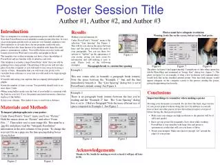
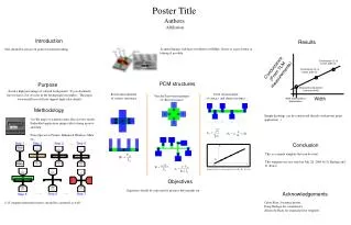
Poster Session Title-Use Upper and Lower Case Authors The University of Texas Health Science Center at San Antonio, San Antonio, TX 78229 ABSTRACT Cell Culture: Cells from a human retinal pigment epithelial cell line (ARPE-19 from American Type Culture Center #CRL 2302, lot # 1263984) were revived in a mixture of 90% 1: 1 DMEM and F-12 medium with 3 mM L-Glutamine, and 10% fetal bovine serum ( FBS ) and incubated at 37oC in 10% CO2. Medium was changed within 24 hr of seeding and then every three days thereafter. ARPE-19 is a spontaneously-arising RPE cell line from the normal eye of a 19 year-old head trauma patient. These cells form stable monolayers and exhibit morphological and functional polarity. They also express the RPE-specific markers CRALBP and RPE 65 (Dunn, et al., Exp. Eye Res. 62: 155-169, 1996). Harvest and Processing of Confluent RPE Cells: Within 24 hr, revived cells adhered to the culture plate (D-150, 30 ml of medium) and changed morphology to an elongated, spindle shape. Cells were near confluency in 5 days from seeding (Fig. 1a) and were harvested on the 7th day (confluent cells, Fig. 1b). After the removal of cell medium by aspiration, cells were rinsed with HBSS and then treated with trypsin/EDTA for detachment. They were then harvested, washed, and centrifuged. Collected cells from three D-150 plates were homogenized in 2 ml of Tris buffer (pH 8) and then centrifuged (100,000 xG) to separate particulate membranes from cytosol. RESULTS Cell Culture: Cells from a human retinal pigment epithelial cell line (ARPE-19 from American Type Culture Center #CRL 2302, lot # 1263984) were revived in a mixture of 90% 1: 1 DMEM and F-12 medium with 3 mM L-Glutamine, and 10% fetal bovine serum ( FBS ) and incubated at 37oC in 10% CO2. Medium was changed within 24 hr of seeding and then every three days thereafter. ARPE-19 is a spontaneously-arising RPE cell line from the normal eye of a 19 year-old head trauma patient. These cells form stable monolayers and exhibit morphological and functional polarity. They also express the RPE-specific markers CRALBP and RPE 65 (Dunn, et al., Exp. Eye Res. 62: 155-169, 1996). Harvest and Processing of Confluent RPE Cells: Within 24 hr, revived cells adhered to the culture plate (D-150, 30 ml of medium) and changed morphology to an elongated, spindle shape. Cells were near confluency in 5 days from seeding (Fig. 1a) and were harvested on the 7th day (confluent cells, Fig. 1b). After the removal of cell medium by aspiration, cells were rinsed with HBSS and then treated with trypsin/EDTA for detachment. They were then harvested, washed, and centrifuged. Collected cells from three D-150 plates were homogenized in 2 ml of Tris buffer (pH 8) and then centrifuged (100,000 xG) to separate particulate membranes from cytosol. RESULTS (cont.) Figure 4 Figure 5 • CONCLUSIONS • 11-cis Retinyl Ester Hydrolase (REH) activity is expressed in the membrane fraction of cultured human retinal pigment epithelial cells, ARPE 19; • Our data demonstrate that ARPE 19 has properties (i.e. 11-cis REH activity) similar to freshly isolated RPE cells; • Therefore, ARPE 19 has functional properties appropriate for in vitro studies of visual cycle physiology of the RPE. • 11-cis Retinyl Ester Hydrolase (REH) activity is expressed in the membrane fraction of cultured human retinal pigment epithelial cells, ARPE 19; • Our data demonstrate that ARPE 19 has properties (i.e. 11-cis REH activity) similar to freshly isolated RPE cells; • Therefore, ARPE 19 has functional properties appropriate for in vitro studies of visual cycle physiology of the RPE. MATERIALS and METHODS Cell Culture: Cells from a human retinal pigment epithelial cell line (ARPE-19 from American Type Culture Center #CRL 2302, lot # 1263984) were revived in a mixture of 90% 1: 1 DMEM and F-12 medium with 3 mM L-Glutamine, and 10% fetal bovine serum ( FBS ) and incubated at 37oC in 10% CO2. Medium was changed within 24 hr of seeding and then every three days thereafter. ARPE-19 is a spontaneously-arising RPE cell line from the normal eye of a 19 year-old head trauma patient. These cells form stable monolayers and exhibit morphological and functional polarity. They also express the RPE-specific markers CRALBP and RPE 65 (Dunn, et al., Exp. Eye Res. 62: 155-169, 1996). Harvest and Processing of Confluent RPE Cells: Within 24 hr, revived cells adhered to the culture plate (D-150, 30 ml of medium) and changed morphology to an elongated, spindle shape. Cells were near confluency in 5 days from seeding (Fig. 1a) and were harvested on the 7th day (confluent cells, Fig. 1b). After the removal of cell medium by aspiration, cells were rinsed with HBSS and then treated with trypsin/EDTA for detachment. They were then harvested, washed, and centrifuged. Collected cells from three D-150 plates were homogenized in 2 ml of Tris buffer (pH 8) and then centrifuged (100,000 xG) to separate particulate membranes from cytosol. Protein Determination and Enzyme Assay: Protein amount in cytosol and membrane fractions were determined by the Bradford Dye Reagent method. The activity of 11-cis REH was determined by a sensitive radiometric method described previously(Mata, et al., J. Biol. Chem. 267: 9794-9799, 1992 and Tsin, et al. Meth. Enzymol. 316: 551-569, 00). Figure 2 Figure 1 REFERENCES 1. 11-cis Retinyl Ester Hydrolase (REH) activity is expressed in the membrane fraction of cultured human retinal pigment epithelial cells, ARPE 19; 2. Our data demonstrate that ARPE 19 has properties (i.e. 11-cis REH activity) similar to freshly isolated RPE cells; 3. Therefore, ARPE 19 has functional properties appropriate for in vitro studies of visual cycle physiology of the RPE. 4. 11-cis Retinyl Ester Hydrolase (REH) activity is expressed in the membrane fraction of cultured human retinal pigment epithelial cells, ARPE 19; Figure 3