Download

1 / 42
590 likes | 1.33k Views
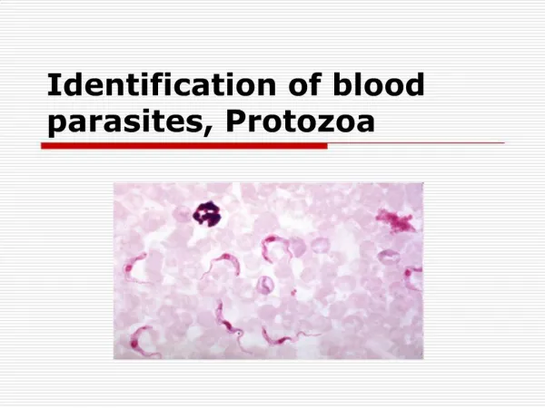
PRACTICAL ON BLOOD PARASITES. Common methods for parasitological diagnosis of malaria. The two methods common in use : 1: Light microscopy 2: Rapid diagnostic tests (RDTs). Laboratory diagnosis of malaria. Rapid diagnostic tests detect malaria antigens.

E N D
Common methods for parasitological diagnosis of malaria The two methods common in use : 1: Light microscopy 2: Rapid diagnostic tests (RDTs).
Laboratory diagnosis of malaria Rapid diagnostic tests detect malaria antigens
Laboratory diagnosis of malaria Light microscopy:1: Preparing blood film
Laboratory diagnosis of malaria Light microscopy:1: Preparing blood film
Laboratory diagnosis of malaria Light microscopy:1: Preparing blood film
Laboratory diagnosis of malaria Light microscopy:1: Preparing blood film
Laboratory diagnosis of malaria Light microscopy:1: Preparing blood film
Laboratory diagnosis of malaria Light microscopy: Thick and thin films
Laboratory diagnosis of malaria CCMOVBD Plasmodium falciparum (trophozoite stage in thin smear) Plasmodium falciparum (trophozoite stage in thick smear)
Trophozoites CCMOVBD CCMOVBD Schizont Gametocyte CCMOVBD CCMOVBD 13 Morphology of Malaria The Malaria Parasite Three developmental stages seen in blood films: • Trophozoite 2. Schizont 3. Gametocyte
CCMOVBD 14 Morphology of Malaria Features of Plasmodium Vacuole Nucleus/chromatin dot Cytoplasm Stippling
Plasmodium vivax Plasmodium falciparum CCMOVBD CCMOVBD Plasmodium malariae Plasmodium ovale CCMOVBD Malaria Tutorials, Wellcome Trust 15 Morphology of Malaria Species of malaria is identified by its microscopic appearance:
Multiple infection Double chromatin CCMOVBD Marginal form 16 Morphology of Malaria Plasmodium falciparum (trophozoite stage) Diagnostic Points: • Small, regular, fine to fleshy cytoplasm • Infected RBCs not enlarged • Numerous, multiple infection is common • Ring, comma, marginal or accole forms are seen; often have double chromatin dots • Maurer’s dots not clearly visible
CCMOVBD 17 Morphology of Malaria Plasmodium falciparum (trophozoite stage in thin smear)
CCMOVBD 18 Morphology of Malaria Plasmodium falciparum (trophozoite stage in thick smear)
CCMOVBD CCMOVBD 19 Morphology of Malaria Plasmodium falciparum (schizont stage) Diagnostic Points: • Small, rarely fill the RBC • Rare in peripheral blood • 16-32 or more merozoites in compact cluster • Single dark pigment • Usually associated with many young ring forms
CCMOVBD 20 Morphology of Malaria Plasmodium falciparum (trophozoite and schizont stages in thin smear)
CCMOVBD CCMOVBD 21 Morphology of Malaria Plasmodium falciparum (gametocyte stage) Diagnostic Points: • Banana-shaped or rounded • Macrogametocyte • small, compact, central chromatin dot • pigments closely adhere to the chromatin • Microgametocyte • broader, shorter and more sausage-shaped • loosely scattered chromatin • golden brown pigments scattered at the central half
CCMOVBD 22 Morphology of Malaria Plasmodium falciparum (gametocyte stage in thin smear)
CCMOVBD CCMOVBD 23 Morphology of Malaria Plasmodium vivax (trophozoite stage) Diagnostic Points: • Infected red cells usually enlarged • Irregular or fragmented cytoplasm (amoeboid) • Mature ring forms tend to be large and coarse • Schuffner’s dots (stippling) are frequently visible
Ring form Developing trophozoite CCMOVBD CCMOVBD CCMOVBD 24 Morphology of Malaria Plasmodium vivax(trophozoite stages in thin smear) Mature Trophozoite
CCMOVBD 25 Morphology of Malaria Plasmodium vivax (schizont stage) Diagnostic Points: • Large, covering almost or the entire enlarged RBC • Few to moderate • 12 – 24 merozoites in irregular cluster • Yellowish-brown loose pigments
Platelet This RBC has two rings. Schüffner’s dots are present in this cell. This RBC is not as enlarged as the others. Schüffner’s dots are not visible. In this malarial infection RBC is enlarged and oval, schuffner's dot is present. Trophozoite ring is 1/3 diameter of cell with heavy chromatin dot
CCMOVBD CCMOVBD CCMOVBD 27 Morphology of Malaria Plasmodium vivax (gametocyte stage) Diagnostic Points: • Round; large • Usually compact nucleus the periphery • Chromatin is deep red or magenta
CCMOVBD 28 Morphology of Malaria Plasmodium vivax (gametocyte stage in thin smear)
CCMOVBD CCMOVBD 29 Morphology of Malaria Plasmodium malariae (trophozoite stage) Diagnostic Points: • Small, few • Ring to rounded, compact, vacuolated or non-vacuolated, • Band forms seen • Dark, scattered pigments • Ziemman’s stipplings not clearly visible • Infected red cell not enlarged
CCMOVBD CCMOVBD 30 Morphology of Malaria Plasmodium malariae (schizont stage) Diagnostic Points: • Small, compact, dark • Usually few • 6-12 merozoites in “rosette” formation, but more often in irregular cluster • Concentrated pigments
Malaria Tutorials, Wellcome Trust 31 Morphology of Malaria Plasmodium ovale Diagnostic Points: • Smaller than P. vivax • Few • Ring to rounded, compact • Single, prominent chromatin • Schizonts with 4-12 merozoites • Reported in the Philippines
CCMOVBD Mixed infections • Infection of an individual by more than one species of Plasmodium • Common in endemic areas in the Philippines • P. falciparum and P. vivax • Can easily be overlooked (blood films must be studied carefully to rule out mixed infections)
CCMOVBD 33 Morphology of Malaria P. vivax trophozoite and P. falciparum gametocyte (Mixed infection, thin smear)
CCMOVBD 34 Morphology of Malaria Plasmodium falciparum
CCMOVBD 35 Morphology of Malaria Plasmodium malariae
36 Morphology of Malaria Plasmodium vivax
37 Morphology of Malaria Plasmodium falciparum
A 25 year-old male from India, who came 3 months ago was admitted in KKUH with a history of severe anaemia and intermittent high grade fever for the last two months not responding to antibiotics. WHAT IS THE DIAGNOSIS? Diagnosis: Plasmodium vivax
A businessman who makes frequent trips to Thailand , presents with intermittent fever . WHAT IS THE DIAGNOSIS? Diagnosis: Plasmodium vivax
A student in KSU who returned three weeks from vacation in Africa , he developed intermittent fever last week and lost consciousness a short time ago. WHAT IS THE DIAGNOSIS? Diagnosis: Plasmodium falciparum
The patient was then treated with schizontocidalantimalarial drugs, a follow-up blood film is shown . ARE THERE ANY PARASITES? WHAT STAGE ? Plasmodium flaciparum , gametocyte stage
A 7 year old child presented with anemia , hepatospenomegaly and fever .Not responding to antimalarials and antibiotics . Bone marrow smear is shown ARE THERE ANY PARASITES? WHAT STAGE ? Leishmania , amastigote stage