Download
1 / 85
850 likes | 995 Views
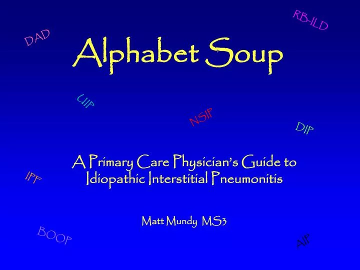
RB-ILD. DAD. Alphabet Soup. UIP. NSIP. DIP. A Primary Care Physician’s Guide to Idiopathic Interstitial Pneumonitis Matt Mundy MS3. IPF. BOOP. AIP. Idiopathic Interstitial Pneumonitis : The Dilemma

E N D
RB-ILD DAD Alphabet Soup UIP NSIP DIP A Primary Care Physician’s Guide to Idiopathic Interstitial Pneumonitis Matt Mundy MS3 IPF BOOP AIP
Idiopathic Interstitial Pneumonitis : The Dilemma Identification of this class of diseases is notoriously difficult. Uncertain etiologies, discrepancies in the descriptions of the clinical, radiographic and pathologic characteristics of the disease processes and the necessity for multidisciplinary communication and cooperation in the clinical investigation all add to the challenge of making an accurate diagnosis. Combined with a seemingly endless string of acronyms used to refer to specific disease subtypes, these factors can make Idiopathic Interstitial Pneumonitis a seemingly insurmountable challenge to clinicians.
So……… • For a Primary Care Physician, what are the most important questions to ask about Idiopathic Interstitial Pneumonitis? • What are the major subtypes of Idiopathic Interstitial Pneumonitis? • What are the important Clinical, Pathologic, and Radiographic findings in each of these subtypes? • What therapies are available when a diagnosis is ultimately made?
What are the most important questions to ask? • By understanding the acronyms and answering just a few questions, a primary care doctor can have good insight into a patients’ disease and its prognosis. This understanding can then be used to guide future therapy and clinical decision making.
Welcome to IIP 101 An quick and easy guide to the basics of Idiopathic Interstitial Pneumonitis A, B, C, D, E, F, G………..
Question #1 What do all the acronyms mean? Clinical Diagnosis Pathologic Diagnosis Idiopathic Pulmonary Fibrosis (IPF)……………………..……..Usual Interstitial Pneumonia (UIP) Desquamative Interstitial Pneumonia (DIP)……………….……………………………….…….….same (DIP) Respiratory Bronchiolitis Interstial Lung Disease (RBILD)…….…………….same (RBILD) Acute Interstitial Pneumonia (AIP)…………………………..…Diffuse Alveolar Damage (DAD) Nonspecific Interstitial Pneumonia (NSIP)………………………………………………………same (NSIP) *adapted from: The American Thoracic Society / European Respiratory Society International Multidisciplinary Consensus Classification 2002
Important clarifications about the acronyms • Each pathologic diagnoses has a distinct pattern • Each of the pathologic patterns has unique and consistent radiographic findings • The pathologic and radiographic patterns can have multiple etiologies • A clinical diagnosis of a corresponding Idiopathic Interstitial Pneumonitis can only be made when no cause for the pathology can be identified
ILD Vs. DLD: What, more acronyms? • Interstitial Lung Disease (ILD), a term used by pathologists refers to the same set of diseases that radiologists call Diffuse Lung disease(DLD). • There are over 150 identified processes that fall under this description • All of them can lead to lung scarring and respiratory failure • Examples include: occupational, exposure related and drug induced diseases, eosinophilic pneumonias, sarcoidosis, collagen vascular related ILD, granulomatous disease and finally Idiopathic Interstitial Pneumonitis
So……… IIP is a subset of ILD
And……….. IPF, DIP, RB-ILD, AIP and NSIP Are all subtypes of IIP
Question #2 What is the most important distinction to make? Everything Else UIP Vs.
Why? Complete recovery is possible with all types of Idiopathic Interstitial Pneumonitis Except UIP/IPF!!!
Because of its poor prognosis and refractoriness to treatment, it is essential to identify cases of usual interstitial pneumonia / idiopathic pulmonary fibrosis • Things to remember: • Immunosuppression or smoking cessation will improve survival all types of IIP EXCEPT UIP/IPF • The median survival after diagnosis for UIP/IPF is 2.5-3.5 years • No therapy has been shown to reduce the severity of pathologic findings or improve the prognosis of UIP/IPF
The diagnosis of UIP/IPF does not require pathologic confirmation • With a high clinical suspicion, the characteristic radiographic findings of UIP are specific enough to be sufficient for diagnosis • Studies show that combining clinical history, including pulmonary function tests, and CT findings alleviates the need for biopsy in up to 89% of patients • When conflicting or inconclusive findings arise, a biopsy specimen can solidify the diagnosis
Again……. • The diagnosis of UIP/IPF implies many things, including: • There is probably no need for a biopsy • Treatment of any sort is likely to have little, if any effect • MOST IMPORTANTLY: UIP/IPF implies a very poor prognosis for the patient no matter what their age, gender or extent of disease
Question #3 What is “ground glass” anyway? *many of the radiographic patterns of IIP involve ground glass opacities, therefore, the ability to identify ground glass on a CXR and CT is essential
Ground Glass Opacity: “focal or diffuse areas of ill-defined, hazy, increased lung attenuation, which cause pulmonary vascular indistinctness, yet through which vessels can still be identified.”* *Nowers et al
Ground Glass Opacity: The ground glass pattern can be identified on both CT and conventional radiographs
Ground Glass Opacity: Ground glass falls on a spectrum of increasing opacification that culminates in consolidation consolidation Ground glass
What are the major subtypes of IIP? and What are the most pertinent clinical, radiographic and pathologic findings? *remember that IIP is a multidisciplinary diagnosis
Important subtypes of IIP • UIP/IPF (usual interstitial pneumonia/idiopathic pulmonary fibrosis) • NSIP (nonspecific interstitial pneumonia) • AIP/DAD (acute interstitial pneumonia/diffuse alveolar damage) • RB-ILD (respiratory bronchiolitis interstitial lung disease) • DIP (desquamative interstitial pneumonia) *RB-ILD and DIP are strongly associated with smoking and respond well to smoking cessation. Therefore, arguments have been made against classifying them as idiopathic.
UIP IPF
UIP / IPF • (Usual Interstitial Pneumonia/Idiopathic Pulmonary Fibrosis) • Clinical Presentation -History • age: usually greater than 50 • male to female ratio: between 1:1 and 2:1 • 75% have a smoking history • insidious exertional dyspnea which is disabling over time • nonproductive cough - refractory to antitussive medication • fever, malaise and arthralgia reported in 50%
UIP / IPF • (Usual Interstitial Pneumonia/Idiopathic Pulmonary Fibrosis) • Clinical Presentation - Physical Exam • tachypnea • bibasilar inspiratory crackles • 75% have digital clubbing • PFTs show restrictive physiology ( VC and TLC, FEV1 to FVC ratio) • reduced diffusion capacity (DLCO) • symptoms usually start >6 months before clinical presentation
UIP / IPF (Usual Interstitial Pneumonia/Idiopathic Pulmonary Fibrosis) Digital Clubbing
UIP / IPF • (Usual Interstitial Pneumonia/Idiopathic Pulmonary Fibrosis) • Differential Diagnosis (etiologies that can cause a UIP pattern) • asbestosis • collagen vascular disease induced interstitial lung disease • drug toxicity • chronic extrinsic allergic alveolitis
UIP / IPF • (Usual Interstitial Pneumonia/Idiopathic Pulmonary Fibrosis) • Radiographic Findings – Chest Radiograph • Peripheral, lace-like areas of fine reticular opacity • Concentrated posteriorly in the bases • Visible fibrosis in costophrenic recesses on lateral CXR • Ground glass opacification of lower lobes
UIP/IPF Peripheral reticular opacity more predominant in the bases
UIP / IPF • (Usual Interstitial Pneumonia/Idiopathic Pulmonary Fibrosis) • Radiographic Findings – CT • Similar to CXR • Fibrosis • Traction bronchiectasis • Architectural distortion • +- honeycombing • Variable ground glass opacity • Concentrated in lower lobes
UIP/IPF Peripheral ground glass No evidence of honeycombing Area of consolidation
UIP/IPF Peripheral traction bronchiectasis and honeycombing More predominant in lung base
UIP/IPF Bilateral peripheral honeycomb cysts
UIP/IPF marked honeycombing – end stage disease
UIP / IPF (Usual Interstitial Pneumonia/Idiopathic Pulmonary Fibrosis) If the clinical and radiographic findings are consistent and suggest UIP/IPF a diagnosis can be made without a biopsy. Pathologic evidence is only necessary in cases where the diagnosis is uncertain *all other forms of IIP require pathologic confirmation for diagnosis
UIP / IPF • (Usual Interstitial Pneumonia/Idiopathic Pulmonary Fibrosis) • Pathologic Findings • Video Assisted Thoroscopic Biopsy (VATS) required because of the peripheral distribution of disease • Patchy, dense subpleural collagenous fibrosis +/- Honeycombing • Fibroblastic foci of acute injury • Very little inflammation • Areas of interspersed normal lung • The hallmark is spatial and temporal heterogeneity of disease
UIP/IPF – gross specimen Small, firm, fibrosed lung seen in advanced disease
UIP/IPF – gross specimen White tissue represents interstitial fibrosis
UIP/IPF – histologic section Spatial heterogeneity: fibrosis with interspersed normal lung
UIP/IPF – histologic section Area of acute lung injury: fibroblastic foci
UIP/IPF – histologic section Honeycombing: the final stage of UIP/IPF
UIP / IPF • (Usual Interstitial Pneumonia/Idiopathic Pulmonary Fibrosis) • Treatment and Prognosis • no response to steroids or other immunosuppressive drugs • smoking cessation does not change course of disease • course marked by periods of rapid clinical deterioration • 5 year survival rate: 10%-50% • median survival after diagnosis: 2.5-3.5 years • prognosis worse in men
NSIP • Nonspecific Interstitial Pneumonia • Clinical Presentation - History and Physical • less chronic than UIP with a duration of months to years • age: 39-50 years • male to female ratio: 2:1 • 68% with smoking history • dyspnea, cough and occasional fever • tachypnea, crackles, no digital clubbing • PFT’s: restrictive
NSIP • Nonspecific Interstitial Pneumonia • Differential Diagnosis (etiologies that can cause an NSIP pattern) • collagen vascular disease induced interstitial lung disease • extrinsic allergic alveolitis • resolving acute lung injury • drug induced pneumonitis • infection (HIV)
NSIP • Nonspecific Interstitial Pneumonia • NSIP is further divided into subtypes • Cellular NSIP is the less severe form that occurs in younger people • Fibrotic NSIP shares many of the features of UIP and has a worse prognosis than the cellular subtype • These distinctions become important for both the radiographic and pathologic investigation of the disease • Although distinctions have been made, it is clear that the two subtypes lie on a continuum and overlap does exist
NSIP • Nonspecific Interstitial Pneumonia • Radiographic Findings – cellular subtype • Relative predominance of ground glass opacity with fewer areas of fibrosis • Can be distributed diffusely or in a basilar predominant pattern • Peribronchovascular thickening and areas of consolidation • Occasional traction bronchiectasis secondary to chronic inflammation
NSIP • Nonspecific Interstitial Pneumonia • Radiographic Findings – fibrotic subtype • Relative predominance of fibrosis with areas of ground glass opacity • Reticular opacities and traction bronchiectasis can be seen • Severe form can be indistinguishable from UIP • Decreased occurrence and severity of honeycombing in the face of severe fibrosis suggests NSIP
NSIP Reticular opacity Bilateral basal ground glass and reticular opacities with peripheral distribution Ground glass
NSIP Reticular opacity Ground glass Traction bronchiectasis