Download
1 / 32
350 likes | 540 Views
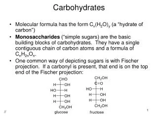
Carbohydrates. Chapter 9 (all of it). Monosaccharide (1) Oligosaccharide (2-20) Polysaccharide (>20). Representative monosaccharides, “simple sugars” single polyhydroxy aldehyde or ketone unit. trioses. hexoses. pentoses in DNA and RNA.

E N D
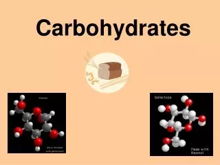
Carbohydrates Chapter 9 (all of it) Monosaccharide (1) Oligosaccharide (2-20) Polysaccharide (>20)
Representative monosaccharides, “simple sugars” single polyhydroxy aldehyde or ketone unit trioses hexoses pentoses in DNA and RNA
D-aldoses D refers to configuration at chiral center most distance from carbonyl carbon you will be responsible for knowing the structures of the sugars named in boxes. These are the most common in nature. (Marked with )
Two sugars that differ only in the configuration around one carbon atom are called epimers D-Glucose and two of its epimers
Pyranoses and furanoses anomers
A chair conformation of Conformations are preferred that have bulky substituents in the equatorial positions
Organisms contain a variety of hexose derivatives R =lactic acid
Aldehydes as reducing agents OH O- H + H2O R-C R-C + 2 H+ + 2 e- = = O O O2 + 2 H+ + 2 e- H2O2 H R-C R-C = = O 1. 2. 3. + O2 + H2O + H2O2 O
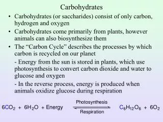
Sugars as reducing agents (Ketoses isomerize to aldoses under the conditions used for these oxidation reactions with copper ions and thus also act as reducing sugars.)
Disaccharides contain an O-glycosidic Bond This ring can’t open up and is no longer reducing This ring can open up to an aldehyde and is still reducing
Starch and Glycogen are energy storage molecules Starch granules in chloroplast Amyloseandamylopectin Glycogen granules in hepatocyte
Starch and Glycogen Short segment of amylose, a long, unbranched polymer of D-glucose residues in (14) linkage A cluster of amylose and amylopectin such as is shown above is believed to occur in starch granuoles. Glycogen is similar to amylopectin, but much more highly branched. Like amylose, amylopectin is also high mw, but it is highly branched. Above is an (16) branch point of amylopectin.
The (14) linkages cause the chains in starch and glycogen to curl up and make the resulting granules very dense.
Cellulose and chitin are structural homopolysaccharides Two units of a cellulose rigid chain. The D-glucose residues are in the (b14) configuration and therefore straight. A short segment of chitin, a homopolymer of N-actyl-D-glucosamine units in (b14) linkage Two parallel cellulose chains, showing inter- and intrastrand hydrogen bonding cross-links between them
Bacterial Cell Walls Contain Peptidoglycans. The rigid component of bacterial cell walls is a heteropolymer of alternating (b14) linked N-acetylglucosamine and N-acetylmuramic acid residues. The linear polymers lie side by side in the cell wall, cross-linked by short peptides, the exact structure of which depends on the bacterial species. Peptidoglycan of the cell wall of Staphylococcus aureus Peptides (strings of colored spheres) covalently link N-acetylmuramic acid residues in a neighboring polysaccharide chains. only in gram-positive bacteria
Repeating units of some common glycosaminoglycans of extracellular matrix. The molecules are copolymers of alternating uronic acid and amino sugar residues, with sulfate esters in any of several positions. The ionized carboxylate and sulfate groups (red) give these polymers their characteristic negative charge. Found in synovial fluid in joints Found in cartilage and tendons Found in horny structures
Proteoglycans: macromolecules of the cell surface or extracellular matrix in which one or more glucosaminoglycan chains are joined covalently to a membrane protein or a secreted protein Act as identity tags, destination labels, mediators of specific cell-cell interactions and interactions between the cell and extracellular matrix Trisaccharide linker
Glycoproteins are information-rich conjugates containing oligosaccharides
Bacterial lipopolysaccharides are dominant surface feature of the outer membrane of gram-negative bacteria. Space-filling model based on crystal structure of lipopolysaccharide from E. coli. (One of the fatty acid acyl chains was not visible.)
Lectins, found in all organisms, are proteins that bind carbohydrates with great affinity and specificity
Roles of oligosaccharides in recognition and adhesion at the cell surface Oligosaccharides with unique structures, components of glycoproteins or glycolipids on the outer surface of plasma membrane,that can interact with lectins. Viruses that infect animal cells bind to glycoproteins on the cell as first step in infection. Bacterial toxins, e.g., cholera or pertussis toxins, bind to surface glycolipid before entering cells. Some bacteria, such as Heliobacter pylori, adhere to then colonize or infect animal cells. Lectins called selectin, such as those of T lymphocytes with the endothelial cells of the capillary wall at an infection site.
Lectin-ligand interactions in controlling movement to the site of an infection or injury Lymphocytes Phagocytes