Download
1 / 9
90 likes | 193 Views
Integrating Nanostructures with Biological Structures Investigators: M. Stroscio, ECE and BioE; M. Dutta, ECE Prime Grant Support: ARO, NSF, AFOSR, SRC, DARPA. Problem Statement and Motivation. Quantum Dot.

E N D
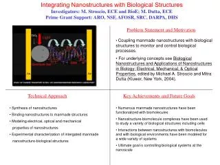
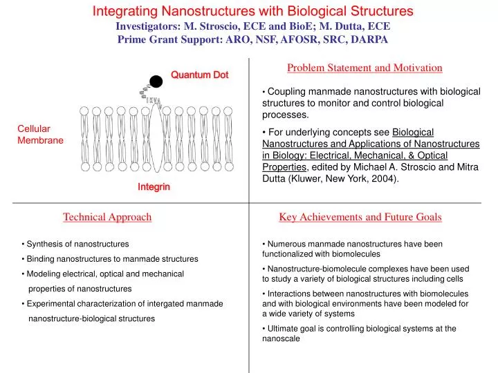
Integrating Nanostructures with Biological Structures Investigators: M. Stroscio, ECE and BioE; M. Dutta, ECE Prime Grant Support: ARO, NSF, AFOSR, SRC, DARPA Problem Statement and Motivation Quantum Dot • Coupling manmade nanostructures with biological structures to monitor and control biological processes. • For underlying concepts see Biological Nanostructures and Applications of Nanostructures in Biology: Electrical, Mechanical, & Optical Properties, edited by Michael A. Stroscio and Mitra Dutta (Kluwer, New York, 2004). Cellular Membrane Integrin Technical Approach Key Achievements and Future Goals • Synthesis of nanostructures • Binding nanostructures to manmade structures • Modeling electrical, optical and mechanical • properties of nanostructures • Experimental characterization of intergated manmade • nanostructure-biological structures • Numerous manmade nanostructures have been functionalized with biomolecules • Nanostructure-biomolecule complexes have been used to study a variety of biological structures including cells • Interactions between nanostructures with biomolecules and with biological environments have been modeled for a wide variety of systems • Ultimate goal is controlling biological systems at the nanoscale
Neurotronic Communication: Electronic Prostheses To Treat Degenerative Eye Disease Investigators: John R. Hetling, Bioengineering Prime Grant Support: The Whitaker Foundation Problem Statement and Motivation • Retinitis Pigmentosa (RP) is a potentially blinding disease for which there are no cures; one in 4000 people are diagnosed with RP • Microelectronic prostheses represent a potential treatment option for RP • Our objective is to learn to stimulate the diseased retina with microelectrodes such that useful information is conveyed to the mind’s eye of the blind patient Key Achievements and Future Goals Technical Approach • This novel approach is the only means to study electrical stimulation of the retina at the cellular level, in vivo, in a clinically-relevant animal model • Using pharmacological dissection, we have begun to identify the types of retinal neurons targeted by electrical stimulation • Ultimate Goal: To communicate the visual scene to the diseased retina with the highest resolution possible • The Goal will be achieved by optimizing the design of the microelectrode array and the stimulus parameters • The response of the retina to electrical stimulation is studied in vivo • Microelectrode arrays, 12 um thick (above, right), are fabricated in the UIC MAL and surgically placed beneath the retina in the eye (above, left) • The response of the retina to electrical stimulation is recorded and compared to the response to natural light stimuli • We use a unique transgenic rat model of retinal degenerative disease developed in our laboratory
Microscopic Magnetic Resonance Elastography Investigators: Richard L. Magin, Bioengineering; Shadi F. Othman, Bioengineering; Thomas J. Royston, Mechanical and Industrial Engineering Prime Grant Support: NIH R21 EB004885-01 Problem Statement and Motivation • Disease changes the mechanical properties of tissues • Palpation by physician requires physical contact • Propose a noninvasive way (MRI) to measure the stiffness of biological tissues (elastography) • Use the elastography system to measure the mechanical properties of regenerating tissue • Extend the technique to high magnetic field systems to allow micoroscopic resolution Three dimensional shear wave through agarose gel Key Achievements and Future Goals Technical Approach • Generate shear waves in the tissue • Apply magnetic resonance imaging (MRI) to capture shear wave motion • Measure the shear wavelength through the sample • Convert the shear wavelength to shear stiffness • Improving elastography resolution to 34 mm x 34 mm for a 500 mm slice • Monitoring the growth of osteogenic tissue engineered constructs • Applying high resolution microelatography in vivo
Biological Signal Detection for Protein Function Prediction Investigators: Yang Dai Prime Grant Support: NSF Text File of Protein description Sequences Problem Statement and Motivation Coding Vectors • High-throughput experiments generate new protein sequences with unknown function prediction • In silico protein function prediction is in need • Protein subcellular localization is a key element in understanding function • Such a prediction can be made based on protein sequences with machine learners • Feature extraction and scalability of learner are keys. MASVQLY ... …HKEPGV Machine Learner specific subcellular and subnuclear localization Key Achievements and Future Goals Technical Approach • Use Fast Fourier Transform to capture long range correlation in protein sequence • Design a class of new kernels to capture subtle similarity between sequences • Use domains and motifs of proteins as coding vectors • Use multi-classification system based on deterministic machine learning approach, such as support vector machine • Use Bayesian probabilistic model • Developed highly sophisticated sequence coding methods • Developed an integrated multi-classification system for protein subcellular localization • Developed a preliminary multi-classification system for subnuclear localization • Will incorporate various knowledge from other databases into the current framework • Will design an integrative system for protein function prediction based on information of protein localizations, gene expression, and protein-protein interactions
Computational Protein Topographics for Health Improvement Jie Liang, Ph.D. Bioengineering Prime Grant Support: National Science Foundation Career Award, National Institutes of Health R01, Office of Naval Research, and the Whitaker Foundation. Protein surface matching Problem Statement and Motivation • The structure of proteins provide rich information about how cells work. With the success of structural genomics, soon we will have all human proteins mapped to structures. • However, we need to develop computational tools to extract information from these structures to understand how cell works and how new diseases can be treated. • Therefore, the development of computational tools for surface matching and for function prediction will open the door for many new development for health improvement. Evolution of function Key Achievements and Future Goals Technical Approach • We have developed a web server CASTP (cast.engr. uic.edu) that identify and measures protein surfaces. It has been used by thousands of scientists world wide. • We have built a protein surface library for >10,000 proteins, and have developed models to characterize cross reactivities of enzymes. • We also developed methods for designing phage library for discovery of peptide drugs. • We have developed methods for predicting structures of beta-barrel membrane proteins. • Future: Understand how protein fold and assemble, and designing method for engineering better proteins and drugs. • We use geometric models and fast algorithm to characterize surface properties of over thirty protein structures. • We develop evolutionary models to understand how proteins overall evolve to acquire different functions using different combination of surface textures. • Efficient search methods and statistical models allow us to identify very similar surfaces on totally different proteins • Probablistc models and sampling techniques help us to understand how protein works to perform their functions.
Structural Bioinformatics Study of Protein Interaction Network Investigators: Hui Lu, Bioengineering Prime Grant Support: NIH, DOL Protein-DNA complex: gene regulation DNA repair cancer treatment drug design gene therapy Problem Statement and Motivation • Protein interacts with other biomolecules to perform a function: DNA/RNA, ligands, drugs, membranes, and other proteins. • A high accuracy prediction of the protein interaction network will provide a global understanding of gene regulation, protein function annotation, and the signaling process. • The understanding and computation of protein-ligand binding have direct impact on drug design. Technical Approach Key Achievements and Future Goals • Data mining protein structures • Molecular Dynamics and Monte Carlo simulations • Machine learning • Phylogenetic analysis of interaction networks • Gene expression data analysis using clustering • Binding affinity calculation using statistical physics • Developed the DNA binding protein and binding site prediction protocols that have the best accuracy available. • Developed transcription factor binding site prediction. • Developed the only protocol that predicts the protein membrane binding behavior. • Will work on drug design based on structural binding. • Will work on the signaling protein binding mechanism. • Will build complete protein-DNA interaction prediction package and a Web server.
Carcinogenic Potential of Wireless Communication Radiation Investigators: James C. Lin, PhD, Electrical and Computer Engineering; and Bioengineering Prime Grant Support: Magnetic Health Science Foundation Problem Statement and Motivation • Wide Spread Use of Cell Phone Technology • Concerns about Health and Safety • Plectin is A High Molecular Weight Protein • Plectin Immunoreactivity Follows Brain Injury • Mutation of Plectin Identified With Signs of Neurodegenerative Disorder Immunolabeling of Irradiated Rat Brain Using Monoclonal Antibody, Pletin. Key Achievements and Future Goals Technical Approach • Irradiate Young Adult Rats (300 g) in Plexiglass Holder • Produce Power Deposition Patterns in Rat Brains Comparable to Those in Humans • Brains Were Removed and Incubated • Floating Sections Were Used for Immunocytochemistry • Use Monoclonal Antibody - plectin - Labeling • Examination by Light Microscopy • Immunolabeling of Irradiated Rat Brain Showed Increased Glial Fibrillary Acidic Protein (IFAP) • GFAP Plays An Important Role in Glial Reactions After Lesions • Preliminary Results Indicate There is No Difference in Expression Pattern of Plectin Among the Brains Tested at Peak SAR levels of 0, 1.6 and 16 W/kg in the brain. • Additional Experiments to Establish Statistical Validity
Engineering Better Brain Implants for the Future of Medicine Patrick J. Rousche, Ph.D. Bioengineering, and co-PI Laxman Saggere, Ph.D. Mechancial Engineering Prime Grant Support: National Science Foundation Career Award and National Institutes of Health R21…> 1 2 3 4 5 6 Microneurosurgery Problem Statement and Motivation Device Manufacture • The complex neural tissue of the brain is the source or destination for almost all motor and sensory information in the human body • Therefore, multi-channel electrode interfaces with the brain hold great potential as a therapeutic tool for a number of clinical conditions such as paralysis, blindness, and deafness • The architecture of the brain presents an incredible biological, chemical and mechanical design challenge for engineers designing such interfaces <Insert some type of visual picture/diagram, etc.> Electrophysiology AnimalBehavior Key Achievements and Future Goals Technical Approach • Bio-inspired design. By incorporating biocompatible materials and biological surface coatings, brain implants capable of long-term survival and function may be possible. ? • Mechanically-compatible design. Further improvements to implant performance may come from the novel use of flexible implant materials. • Flexible, biocompatible, electrode arrays are developed in the MAL and tested in a rat model. • Neural cell culture is also used in the initial design phase to better understand the interactions at the neuron-device interface. • Development of a cell-culture test chamber • Demonstration of sensory and motor brain signal recording in awake and behaving rats • Beginning of a related study to study stroke in collaboration with the UIC Department of Neurosurgery • Extension of the animal work into bio-robotics • Presentations at IEEE-EMBS (Engineering in Medicine and Biology) conferences • Future: Engineering analysis and design study for optimization of an electrode design suitable for human auditory cortex to treat deafness in humans
Development of a Functional Optical Imaging (FOI) Technique for Studying Retina Investigators: David M. Schneeweis,BioE Prime Grant Support: Pending Problem Statement and Motivation • A noninvasive, high throughput method is required to study the patterns of electrical activity in large numbers of nerve cells in the retina • This is critical for understanding retinal function in normal and diseased retina, and for evaluating retinal prostheses and other therapies for treating blindness • Optical methods offer certain key advantages over classical electrode recording techniques that are labor intensive, invasive, and yield information about only one or a small number of cells at a time Multi-photon microscopy images of isolated rat retina. Each image is at a different layer. Cell membranes are labeled with a fluorescent VSD, and appear bright. Key Achievements and Future Goals Technical Approach • Protocols have been established for loading a particular VSD into cell membranes • The entire thickness of the retina can be imaged with single cell resolution (see figure) • Parameters for imaging the VSD using MPM have been established • Small changes in fluorescence of the VSD can be measured with suitable speed and resolution • Future goals include demonstrating that FOI can measure physiologically relevant voltage changes, and using FOI to study visually or electrically evoked signals in isolated retina of rat • Key elements in Functional Optical Imaging (FOI): • Voltage sensitive dyes (VSDs) are fluorescent molecules that can be delivered to cell membranes, as shown above for a rat retina • Changes in cell voltage cause changes in the optical properties of VSDs • Multi-photon microscopy (MPM) is a technique that allows high resolution imaging of thicker tissues, such as retina • MPM combined with VSDs offers the promise of simultaneously studying the functional electrical activity of large numbers of retinal cells