Download
1 / 20
200 likes | 349 Views
Lehninger Ch. 16 BIO 322 Recitation 3 / Spring 2013. The Citri c Acid Cycle. Glycolysis – complete oxidation of glucose Respiration - pyruvate to water and carbondioxide Cellular Respiration occurs in three major stages.

E N D
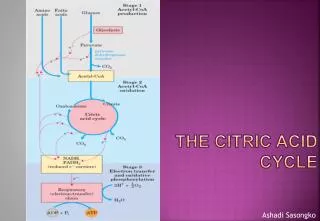
Lehninger Ch. 16 BIO 322 Recitation 3 / Spring 2013 TheCitric AcidCycle
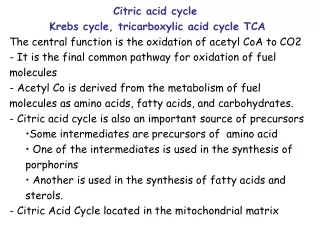
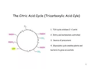
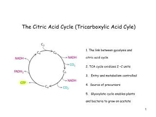
Glycolysis – complete oxidation of glucose • Respiration - pyruvate to water and carbondioxide • Cellular Respiration occurs in three major stages. • 1) Glucose,FA, AA – oxidized to yield 2C fragments in the acetyl group of acetyl-CoA. • 2) Acetyl groups are fed in to TCA cycle, enzymatically oxidizes them to CO2. Energy conserved as NADH and FADH2. • 3) NADH and FADH2 are oxidized to give up protons (H+) and electrons. Electrons are transfered to O2 via ETC. (Release of large amount of ATP via oxidative phosphorylation)
Pyruvate Dehydrogenase Complex • 5 cofactors, 4 of them are vitamins • Covalent modification and allosteric regulation example • Similarity in structure, cofactor req., rxn mech. to • α-ketoglutarate dehydrogenase of TCA cycle • Branched chain α-keto acid dehydrogenase, from oxidative pathway of several AA. Oxidative decarboxylation – irreversible oxidation process, carboxyl group from pyruvate is removed from as CO2. NADH to ETC (Hydride ion – 2 electrons to oxygen) – 2,5 ATP per NADH
TPP (Thiamine pyrophophate) – thiamine (B1) • FAD (Flavin adenine dinucleotide) – riboflavin (B2) • Coenzyme A (CoA, CoA-SH) – pantothenate (B5) • NAD (Nicotinamide adenine dinucleotide) – niacin (B3) • Lipoate • CoA – thiol (-SH) group – acyl carrier • Acyl forms covalent linkage to thiol group • (High standard free energy of hydrolysis) – transfer potential • Lipoate – two thiol groups – reversible oxidation to disulphide linkage • Can undergo redox rxns – electron and acyl carrier.
Identical rxn to pyruvate decarboxylase (Pyruvate to oxalaacetate), C1 of pyruvate to CO2. C2 of pyruvate in the aldehyde form to TPP as hydroxyethyl group. Rate limiting and Substrate Specificity. • Hydroxyethyl group into carboxylic acid (acetate) form. 2 electrons removed in this reaction reduce –S-S of lipoyl group of E2 to two thiol (-SH) groups. • Acetyl group from above redox, esterified first to one lipoyl-SH group, then transesterified to CoA to form acetyl-CoA. • 4) Regeneration oxidized of disulphide form of lipoyl group in E2.
2e + acetyl from pyruvate in E1 to lipollysyl arms of E2, transferring them to E3. Substrate chaneling – intermediates never leave the complex, substrate of E2 is kept at high conc., protects acetyl group to be used by other enzymes. Thiamine deficiency – Beriberi – neuronal dysfunction (white rice, large amounts of alcohol Consumption, high pyruvate concentration in the blood.
Citrosysl CoA – transient intermediate • Free CoA and citrate as products • The large negative free energy is essential to the operation TCA cycle because the concentration of oxaloacetate is normally very low. • CoA sent back to PDH.
Reversible – cis-aconitate intermediate • Addition of water • At pH 7.4, 25˚C the eq. mix contains less 10% isocitrate. • However in the cell, because isocitrate is rapidly consumed in the next step of the TCA cycle, isocitrate formation is favored. • Contains iron-sulphur center (iron is used for heme, cytochromes, Fe-S proteins) • Transferrin – iron transporter • Ferritin – iron storage • When cells are depleted of iron, acotinase loses its activity , apo-acotinase binds specific mRNAs for transferrin receptor and ferritin. • Regulating protein synthesis at translational level.
Oxidative decarboxylation • NAD+ and NADP+ requiring enzymes
Identical Rxn to PDH • E1 is structurally similar but AA sequences differ, thus they have different binding specifity. • PDH – pyruvate • α-ketoglutarate dehydrogenase – α-ketoglutarate • E2 – similar both have lipoyl moieties. • E3 – identical • CoA as the carrier of succinyl group
Enzyme phosphorylated at a His residue in the active site. • This phosphoryl group is transferred to ADP or GDP, dependent on the type of isozymes. • Stabilized active site (phosphoenzyme intermediate) by two subunits oriented in such a way that they confer a partial positive charge on the negatively charged phospho-His residue. • Formation of ATP at the expense of energy released by oxidative carboxylation is substrate level phosphorylation
Mitoch. inner memb. 3 FE-S and FAD – electrons pass before these ETC. 1,5 ATP molecules per electrons (respiration linked phosphorylation)
Because oxaloacetate is removed by step 1, the concentration of oxaloacetate is kept very low. • This condition pulls malate dehydrogenase rxn to the formation of oxaloacetate.
Supply NADH and FADH2 to ETC for oxidative phosphorylation • 2 e from NADH to oxygen – 2,5 ATP • 2 e from FADH2 to oxygen – 1,5 ATP • 32 ATP per glucose molecule • 32 x 30,5 kj/mol = 976 kj/mol • 34% of theoretical max • When corrected for actual free energies, the process is 65% efficient.
Some modern microorganisms lack α-ketoglutarate dehydrogenase • They have reversible enzymes to carry out from oxaloacetate to succinyl-CoA ( reverse of the cycle) • Fermentation because NADH from isocitrate oxidation is recycled to NAD+ by reduction of oxaloacetate to succinate.
Removed intermediates for biosyn are replenished by anaplerotic rxns.
Allosteric inhibiton of PDH by long chains fatty acids, ATP/ADP, NADH/NAD, acetyl-CoA/CoA Allosteric activation of PDH by AMP, CoA, NAD+ Covalent protein modification – PDH inhibited by phosphorylation of E1 at Ser residue on one of the subunits. This kinase is allosterically regulated by ATP levels.
Flow of metabolites in TCA cycle 3 Factors: • Substrate Availability • Inhibition by accumulating products • Allosteric feedback inhibition that catalyze early steps of the cycle


![Tricarboxylic acid cycle (TCA Cycle) [Kreb’s cycle] [Citric acid cycle]](https://cdn3.slideserve.com/6696193/tricarboxylic-acid-cycle-tca-cycle-kreb-s-cycle-citric-acid-cycle-dt.jpg)