Download
1 / 54
540 likes | 639 Views
Molecular Biology of Tumours 2. Dr Orla Sheils. Introduction. All features and properties of individual cells, and ultimately the organisms composed of them are a function of the genes expressed by those cells. Background. Genes of human somatic cells reside in 46 chromosomes.

E N D
Molecular Biology of Tumours 2 Dr Orla Sheils
Introduction • All features and properties of individual cells, and ultimately the organisms composed of them are a function of the genes expressed by those cells.
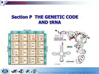
Background • Genes of human somatic cells reside in 46 chromosomes. • 22 pairs of autosomes and 2 sex chromosomes. • Genes consist of stretches of DNA encoding the synthetic function for amino acid chains representing the polypeptide product.
Introduction • Carcinogenesis is a multi step process that leads to uncontrolled growth of cells with unchecked potential for proliferation. • Cancer is caused by the activation or amplification of oncogenes, or by the deactivation or loss of genes that protect against cancer, such as tumour suppressor genes and DNA repair genes.
Introduction • Diverse group of tumours. • Form a spectrum of disease • Clonal malignancies • Characteristic aspects are related to molecular biology of normal B-cells
Classification of lymphomas • Historically lacked precision and accuracy • Potentially compromising diagnosis and therapy • Characterisation of key oncogenic events increased understanding • Combination of molecular and conventional diagnostic tools = optimal
Classification of lymphomas • 25 different lymphoma classification schemes in past 80 years • Initial schemes exclusively morphological • Small vs large cell • Nodular vs diffuse • These criteria remain germaine to contemporary classification –but as components
Introduction • Last two decades of 20th century • First real insights into fundamental biological mechanisms involved in development of human cancer. • Discoveries are based on rapid developments in field of molecular biology.
Antigen Receptor gene rearrangements • Most general diagnostic marker in lymphoma. • Relevant to all lymphocytic cancers. • Antigen receptor genes encode polypeptide units that make up immunoglobulins and T cell receptors. • These are structurally and functionally homologous glycoproteins. • Mediate recognition of antigens and account for the specificity of normal immune response.
B-cells and Immunoglobulin Gene Rearrangement. • B-cells normally produce antibodies • heterodimeric proteins with 2 identical heavy chains, and 2 identical light chains
Immunoglobulins • Ig proteins must be capable of reacting with an infinite number of potential foreign antigens. • need to minimise the amount of info. required at germline level. • recombination • somatic mutation
Immunoglobulins • The three immunoglobulin gene loci IgH, Igk and Igl are located on chromosomes 14q32, 2p12 and 22q11. • All three have the same basic structure. • Gene segments - variable(V), diversity(D), joining(J) • Segments brought together by genetic recombination.
Antigen Receptor Structure • Gathered into three main sets: • V variable • J joining • C constant • IgH gene and b and d T-cell receptor genes also have D (diversity) segment.
Antigen Receptor Structure • Collectively V,D and J segments contain the repertoire of sequence information for the synthesis of all possible variable regions within expressed antigen receptor subunits.
Errors in rearrangement areLymphomagenic • aberrant joinings can occur between immunoglobulin gene segments and some other cellular gene, that confer a survival on the cell. • Burkitt’s Lymphoma - c-myc • Follicular Lymphoma - bcl-2
Burkitt’s Lymphoma • C-myc oncogene (8q24) rearranges into one of the Ig gene loci. • Gene becomes aberrantly expressed as a result of the rearrangement • Continuous proliferative stimulus to the cell.
Follicular Lymphoma • Most common indolent form of lymphoma in Western Hemisphere
Follicular Lymphoma • bcl-2 gene 18q21 is aberrantly translocated into Ig gene locus, leading to inappropriate expression. • bcl-2 provides a growth advantage by extending the lifespan of the cell through its anti-apoptotic function. • Geographical variation in frequency of t14:18 (lower in Asian than North America) • ?environmental effect rather than genetic
Follicular Lymphoma and bcl-2 • about 50% non-Hodgkins lymphoma • generally low grade • 90% have reciprocal translocation between chromosomes 14q32 and 18q21. • Juxtaposition of bcl-2 oncogene to an Ig joining gene. • over-expression of bcl-2 gene product due to juxtaposed Ig and enhancer sequences. • inhibits apoptosis
Follicular Lymphoma and bcl-2 • Chromosome breakpoints are clustered • Facilitates PCR based detection using genomic junctional sequences. • bcl-2 protein localises to membrane systems of nuclei, mitochondria and ER • Provides a survival advantage by inhibiting programmed cell death.
Mantle Cell Lymphoma and bcl-1 • Lymphocytic lymphoma of intermediate differentiation. • Characterised by t(11;14)(q13;q32) • Bcl-1 encodes cyclin-D • cell cycle regulator involved in progression through G1 phase of cell cycle.
Mantle Cell Lymphoma and bcl-1 • Can operate with other oncogenes to transform cells in-vitro. • Several minor breakpoints have been identified on 11q13. • Using a panel of probes it is possible to detect bcl-1 rearrangements in >70% of MCL. • Other genetic markers: • Deletions of INK4a/ARF locus (p16) => p53 and Rb • Disablement of p27
Diffuse Large Cell Lymphoma - bcl-2 and bcl-6 • Heterogeneous group of lymphomas. • Generally have an aggressive clinical course. • Most frequent abnormalities: • t(14;18)(q32;21) • t(3q27) • ~1/4 have t14:18 and bcl-2 rearrangement.
Diffuse Large Cell Lymphoma - bcl-2 and bcl-6 • Molecular cloning of breakpoints have identified the gene bcl-6 - translocated into IgH gene (22q11). • Translocations may also involve other non-immunoglobulin genes on other chromosomes. • Normal function of bcl-6 – unknown. • Translocation results in deregulation and over-expression of bcl-6 • ~5-10% have c-myc involvement +/- EBV • As these tumours share a common molecular pathogenesis with BL ? variant of BL
Small non-cleaved cell lymphoma, Burkitt’s Type and c-myc. • BL is highly aggressive • association with EBV • Association with unique chromosomal translocation involving c-myc- t(8;14)(q24;q32) and Ig gene locus. • ~85% have t(8;14)(q24;q32) • Remainder have t(2;8)(p12;q24) or t(8;22)(q24;q11) involving Igk or Igl gene loci. • Tumour Suppressor genes – p53
T cell receptors • Expressed by T lymphocytes • Membrane bound heterodimers • ab or gd chains • Variable region – differs widely between receptors produced by different lymphocytes.
T-Cell Lymphoma • Majority involve 14q11 • analogy between Ig genes and B-cell malignancy and TCR and T-cell Lymphoma • Trisomy 3,5,7,19
T-Cell Lymphoma • TCR genes organised similarly and rearranged during maturation • t(10;14)(q24;q11) • c-myc - TCR ad elements
c-myc Fusion Transcript • t(2;8)(q34;q24) • increased stability of c-myc
Chromosome translocations • Common in lymphoma • Oncogenes that are modified in structure or expression due to the translocation process are implicated in malignant behaviour of cells harbouring them. • Good markers for malignancy • Constant through course of disease.
Viral genomes • EBV • (Epstein-Barr Virus) • HTLV-1 • (human t-cell leukaemia/lymphoma virus –1) • HHV8 • (human Herpesvirus type 8)
EBV • Maintained in nuclei of latently infected cells as extracellular DNA circles / episomes • Episomes express: • 5 genes for nuclear antigens (EBNAs). • Latent membrane protein (LMP1) – promotes cellular proliferation.
HTLV-1 • 8.6 kb • 5 genes • Oncogenic mechanism not well understood • After infection RNA genome is reverse transcribed to ds DNA /provirus • Inserts at random points into host genome. • Expresses viral proteins for life of cell.
HTLV-1 • Tax protein interacts with cellular transcription factors e.g. IL2 and IL2r • Stimulates proliferation in infected cells.
HHV8 • Discovered by analysis of KS biopsies. • Body cavity based lymphoma • Castleman’s Disease • ?? Multiple Myeloma
Molecular Pathology Thyroid Neoplasia Papillary Thyroid Cancer
Background • Thyroid Cancer • most frequently occurring endocrine malignancy, • sub-divided into a number of diagnostic /morphological categories. • Papillary thyroid carcinoma (PTC) • Most common thyroid malignancy • Ireland<100 cases/yr, U.S.~20,000 cases/yr • Incidence on the rise - global estimate 0.5 million new cases this year • Fastest growing cancer in women worldwide
Introduction • In relation to thyroid carcinoma, particularly PTC, ionizing radiation is the most notorious initiator of carcinogenesis. • In general, radiation is more likely to induce DNA strand breaks rather than point mutations.
Introduction • When DNA strand breaks occur, especially double stranded breaks, the ability to fully repair the sequence is limited and the consequences are chromosomal rearrangements such as inversions, translocations, gains and deletions. • If these consequences are non-lethal for the cell and cannot be otherwise reversed, they may lead to malignant transformation via altered gene expression, formation of chimeric genes, or loss of tumour suppressor gene function.
Papillary Thyroid Carcinoma Follicular Carcinoma Pathological Pathways ret/BRAF Follicular Epithelial Cell RAS
Papillary Thyroid Cancer Normal thyroid PTC • PTC Variants: • Follicular Variant PTC • Tall Cell Variant PTC • Solid PTC • Solid Trabecular PTC
Existing markers of PTC • ret/PTC • To date 15 chimeric mRNAs involving 10 different genes have been described • Ret/PTC-1 and ret/PTC-3 are the most common types, accounting for 90%. • Morphological variants are likely to reflect variations in tumour biology which have yet to be fully defined.
ret/PTC-1 expression • ret/PTC-1 • Inflammatory response • Associated with altered expression of CAM • ? Response to oxidative stress • Classical variant PTC
ret/PTC-1 expression and associated thryoiditis. Number of cases • Risk factors for autoimmune diseases are genetically linked to the presence of specific Class I or Class II HLA alleles. • Viruses implicated in triggering HT –Coxsackie virus
ret/PTC-3 activation • commonly seen in children exposed to ionizing radiation. • ? Radiation signature • Literature demonstrates correlation with solid/follicular variant morphology, poorer prognosis and aggressive tumor behavior. • Correlation between tumor morphology and specific ret rearrangements
Ret activation and morphology • In the setting of radiation induced PTC it is apparent that specific ret/PTC rearrangements are associated with specific tumour morphology • ret/PTC-1 associated with classic morphology • ?low dose/long latency • ret/PTC-3 associated with solid/follicular morphology and adverse prognosis. • ?higher dose/short latency
BRAF • Raf kinases • Serine/threonine kinases • Function in Ras/Raf/MEK/ERK pathway • 3 isoforms: A-Braf, B-Raf, C-Raf/Raf-1 • BRAF implicated in many cancers
Hierarchical clustering– thyroid resectionsBRAF mut vs BRAF wt PTC • Hierarchical clustering of 1139 genes after t-test (p<0.05). • The column dendrogram clearly shows BRAF mut cases clustering to the right with BRAF wt cases in a distinct left-sided cluster. • Red denotes genes with relative increased expression and green denotes genes with relative decreased expression.