Download
1 / 41
550 likes | 1.37k Views
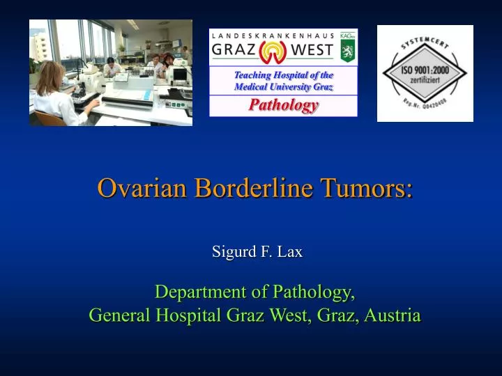
Teaching Hospital of the Medical University Graz. Pathology. Ovarian Borderline Tumors:. Sigurd F. Lax. Department of Pathology, General Hospital Graz West, Graz, Austria. Borderline Tumors of the Ovary 2009: Terminology . Still in debate : Borderline tumor or

E N D
Teaching Hospital of the Medical University Graz Pathology Ovarian Borderline Tumors: Sigurd F. Lax Department of Pathology, General Hospital Graz West, Graz, Austria
Borderline Tumors of the Ovary 2009: Terminology • Still in debate : • Borderline tumor or • Atypically proliferating tumor • Avoid carcinoma of low malignant potential!!
Borderline Tumors benign malignant „borderline“ benign malignant „borderland“ DW Lawrence, IJGP
Distribution of epithelial ovarian tumorsKurman et al. 2004: Blaustein‘s Pathology of the Female Genital Tract
Pathogenetic model of ovarian carcinoma Type I Pathway: low grade serous, mucinous, endometrioid carcinomas K-RAS MSI Low grade carcinoma Borderline-tumor B-RAF PTEN ß-catenin p53 Cystadenoma p53 High grade carcinoma de novo multiple LOH & chromosomal alterations Type II Pathway: high grade serous carcinomas
Borderline tumors: Important questions • Are all BLT potentially malignant? • Can the biological behavior be concluded from histology or from additional techniques? • Published data in the literature: • Heterogeneous study populations and therapy • Answers by the Ovarian Borderline Tumor Workshop (Bethesda 2003: Human Pathology 35 (8), 2004)
Serous Borderline Tumors: Diagnostic Problems • Classification (Definition) • Micropapillary morphology • Invasion • Extraovarian spread/ Implants • Lymph Node Involvement • Staging
Borderline Tumors of the Ovary • Definition WHO 2003 (for serous borderline tumors): • Ovarian tumors of low malignant potential, that show an atypical proliferation of serous cell type, stronger compared to its benign counterpart but without destructive stromal invasion.
Borderline tumors Diagnostic criteria • Cellular atypia • Proliferation / epithelial budding • Detachment of epithelial clusters • Mitoses • Absence of invasion
The low end of borderline morphology: Does it exist? • Quantitative 10% cut-off in the older literature • Considered by most participants of the Bethesda Workshop • Little experience on small borderline foci in benign tumors • Collection of data important for studies • Borderline component <10% influences frozen section accuracy(Brun et al AJOG 2008)
Borderline pattern of a serous carcinoma • High grade nuclear atypia excludes a borderline tumor
Micropapillary pattern in serous BLT Burks et al., AJSP 1997 • Predominance of epithelial proliferation • Fine papillae on a thick fibrous core („Caput medusae“) • Cribriform pattern • Bland cytology with few mitoses but increased N/C ratio • At least 5mm extension
Micropapillary serous borderline tumor Slomovitz et al. AJSP 2002, Prat AJSP 2002,Longacre et al. AJSP 2005 • Frequently bilateral • Frequently higher stages (>I) • Frequently exophytic • Frequently invasive • Frequently associated with invasive implants • But: Prognosis influenced by stage / implant type
Microinvasion in Borderline Tumors • Maximum dimension (WHO): 10 mm2 • Impact of microinvasion on survival • (old data: no influence on prognosis) • Recent data (Longacre AJSP 2005): shorter survival, independent of stage, micropapillary architecture and type of implant
Serous borderline tumors • Impact of extraovarian lesions
Implants • Non- invasive = reactive • Invasive = low grade carcinoma • Indeterminate • Determine higher stage! • Important for prognosis! • Frequently associated with exophytic growth of BLT
Pathogenesis of invasive implants Focal malignant transformation Metastatic spread Implantation of malignant cells Exfoliation Malignant transformation after implantation Borderline Tumor „Field effect“ Independent development of a malignant cell clone Modified acc. to Sherman et al. Hum Pathol 35: 968, 2004
Invasive Implants • Unfavorable prognostic factor for disease-free and overall survival • Prognosis significantly worse compared to non-invasive implants (Bell, Gilks, Prat, Morice)
Long term prognosis based on implant type N=137 Prat and deNictolis AJSP 26: 1111-1128, 2002
Autoimplants • Non-invasive implants on the ovarian surface • Associated with high stage („marker“) • Per se no influence on survival • Cave: no invasion!
Serous BLT and lymph node status • Lymph nodes involved in 30% of BLT • Various patterns of involvement (sinus, infiltration) • No influence on survival • Cave: Endosalpingiosis is no metastasis (in 5-25% of extraovarian malignancies)
Mucinous Borderline Tumors: Diagnostic Problems • Classification (Definition) • Invasion • Origin: Primary versus Metastasis (Differential Diagnosis)
Mucinous borderline tumors • Intestinal type most frequent (85%) • Most stage I and unilateral • Recurrence rare, also in case of intraepithelial carcinoma (IEC) • Pseudomyxoma peritonei (PP) rare (most metastatic) • Endocervical type: no PP; higher stage (implants in 20%) • Important is exclusion of: • Metastasis • Microinvasion > 5mm
Invasion pattern of mucinous ovarian tumors • Infiltrative (desmoplastische Stromareaktion) • Confluent (glandular, cribriform) • Most frequent with mucinous Borderline Tumors • Nodular • Typical for metastases (pankreas, colon, upper GI tract, biliary system)
Tools for the exclusion of metastases • Size (primary carcinoma >12 cm) • Laterality (50% of metastases bilateral) • Immunohistochemistry (CK7, CK20, cdx2, CA125, CA19-9, HAM56) CK20
Endometrioid Borderline Tumors • Rare and infrequent compared to endometrioid carcinomas • Almost all stage I, prognosis excellent • Microinvasion infrequent and of no prognostic impact • Simultaneous endometrial atypical hyperplasia/ carcinoma in 10% • No importance of extraovarian lesions Läsionen • Diagnostic Problem: Invasion (confluent, glandular, expansile)
Problem intraoperative frozen section?Twaalhoven 1999,Houck 2000, Tempfer 2007, Brun 2008 • Accuracy for malignancy of epithelial OvTu 90-94% • Accuracy for borderline tumors approx. 60-70% • Factors: • % Borderlinekomponente (10) • Type (muc), Size of tumor (10/3) • Experience of Pathologist • Additional: size of invasive component
Take home message • Serous borderline tumors: • Be aware of micropapillary architecture, invasion, invasive implants • Staging and careful sampling important • Mucinous borderline tumors: • Rule out metastases from the GI tract
Proposed New Classification of Serous Tumors Kurman & Shih • Serous cystadenoma/adenofibroma • Atypical proliferative serous tumor • Non-invasive low grade MP serous carcinoma • Invasive low grade MP serous carcinoma • High grade serous carcinoma
Current Non-invasive implant Invasive implant Proposed Implant Low grade serous carcinoma Proposed Implant Terminology Kurman & Shih
Proposed New Classification of Mucinous Tumors Kurman & Ronnett • Mucinous cystadenoma/adenofibroma • Atypical proliferative mucinous tumor • Atypical proliferative mucinous tumor with intraepithelial carcinoma • Mucinous adenocarcinoma
Mucinous ovarian tumors: Important gross and microscopic features Riopel et al. AJSP 23:1999
Borderline Tumors of the ovaryKaern et al. Cancer 1992 • 370 patients: 178 mucinous, 174 serous BLT • Follow up > 10 years • Recurrence: 10% (mucinous) / 4% (serous) • Survival: 100% for stage I, 60% for stage III • Prognostic factors: Stage, age, histologic type (serous>non-serous) • PP in 30 cases (12 DOD)
Long term prognosis based on implant type N=80 P<0.002 Morice et al. Ann Oncol 14: 592-8, 2003
Ovarian Borderline Tumor Workshop • Bethesda, USA, 27./28. 8. 2003 • Organized by NIH / NCI • Committee (Kurman, Silverberg, Young, Silva) • 39 participants • Focused on serous and mucinous BLT • Published in: Hum Pathol 35 (8), 2004
Summary BLT Workshop I • Necessity separating an intermediate category • Importance of exact histopathological analysis • Specifications for publications (cell type, unfavorable cases) • „recurrence of borderline tumor“ not acceptable term (specification of lesions necessary; comparison with primary lesions; Cave endosalpingiosis, implants) • Small borderline foci in benign tumors: size important; little experience
Summary of BLT Workshop II • Serous BLT at stages Ia/Ib with or without microinvasion show excellent prognosis (100% 5-yr survival, almost no recurrences) • Separation of micropapillary BLT • Low risk of intracystic BLT, high risk of exophytic micropapillary BLT • „Implants“: limited experience; excellent prognosis of non-invasive implants • Mucinous BLT of gastro-intestinal type always benign (exclusion of cases with pseudomyxoma peritonei and of metastases)
Summary of BLT Workshop III • „sero-mucinous“ BLT similar to serous BLT; limited experience; most benign • Mucinous BLT with intraepithelial carcinoma: Atypia single criterium; Invasion needs to be excluded • BLT with microinvasion: emphasis on BLT; no consensus about size of invasion (5mm??!) • Assessment of architectural features for invasion for mucinous BLT more difficult compared to serous BLT (confluent growth) • Consensus that mucinous tumors with pseudomyxoma peritonei origin from GI-tract (most Appendix, pancreato-biliary system)