Download
1 / 1
10 likes | 109 Views
extremum. n b d b. n f d f. R 2 ’. I. R 2. =2nd. R 1. Y. Y. Y. Y. Y. Y. Incubating. Rinsing. Add BSA. Recognition. An application of Reflectometry Interference Spectroscopy in clinical detection of HBsAg

E N D
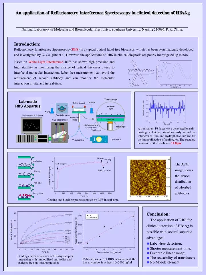
extremum. nb db nf df R2’ I R2 =2nd R1 Y Y Y Y Y Y Incubating Rinsing Add BSA Recognition An application of Reflectometry Interference Spectroscopy in clinical detection of HBsAg National Laboratory of Molecular and Biomolecular Electronics, Southeast University, Nanjing 210096, P. R. China, Introduction: Reflectometry Interference Spectroscopy(RIfS) is a typical optical label-free biosensor, which has been systematically developed and investigated by G. Gauglitz et al. However, the applications of RIfS in clinical diagnosis are poorly investigated up to now. Based on White-Light Interference, RIfS has shown high precision and high stability in monitoring the change of optical thickness owing to interfacial molecular interaction. Label-free measurement can avoid the requirement of second antibody and can monitor the molecular interaction in-situ and in real-time. Transducer Lab-made RIfS Appartus Sample Teflon flow cell Antibody layer Waste Peristaltic pump PC Computer & Software Reflective Probe CCD spectrometer substrate Interference layer (polystyrene) Matching oil A transparent PS layer were generated by spin-coating technique, simultaneously served as interference film and hydrophobic surface for the immobilization of antibodies. The standard deviation of the baseline is 17.8pm. Ta2O5 layer Light source “Y” shape fiber The AFM image shows the dense distribution of adsorbed antibodies Coating and blocking process studied by RIfS in real-time. • Conclusion: • The application of RIfS for clinical detection of HBsAg is possible with several superior advantages: • Label-free detection; • Shorter measurement time; • Favorable linear range; • The reusability of transducer; • No Mobile element. Binding curves of a series of HBsAg samples interacting with immobilized antibodies and analyzed by non-linear regression Calibration curve of RIfS measurement, the linear window is at least 10~5000 ng/ml