Download
1 / 25
310 likes | 423 Views
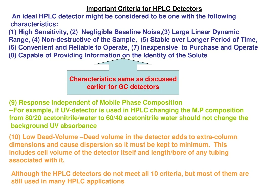
Important Criteria for HPLC Detectors An ideal HPLC detector might be considered to be one with the following characteristics: High Sensitivity, (2) Negligible Baseline Noise,(3) Large Linear Dynamic Range, (4) Non-destructive of the Sample, (5) Stable over Longer Period of Time,

E N D
Important Criteria for HPLC Detectors An ideal HPLC detector might be considered to be one with the following characteristics: • High Sensitivity, (2) Negligible Baseline Noise,(3) Large Linear Dynamic Range, (4) Non-destructive of the Sample, (5) Stable over Longer Period of Time, (6) Convenient and Reliable to Operate, (7) Inexpensive to Purchase and Operate (8) Capable of Providing Information on the Identity of the Solute Characteristics same as discussed earlier for GC detectors (9) Response Independent of Mobile Phase Composition --For example, if UV-detector is used in HPLC changing the M.P composition from 80/20 acetonitrile/water to 60/40 acetonitrile water should not change the background UV absorbance (10) Low Dead-Volume –Dead volume in the detector adds to extra-column dimensions and cause dispersion so it must be kept to minimum. This includes cell volume of the detector itself and length/bore of any tubing associated with it. Although the HPLC detectors do not meet all 10 criteria, but most of them are still used in many HPLC applications
Fixed l Variable l UV-Vis Photodiode Array Amperometry Pulse Amperometry Electrochemical Solute Property Fluorescence Voltammetry Coulometry Refractive Index Bulk Property Deflectance Type Reflectance Type Conductivity Suppressed Non-suppressed Major Types of HPLC Detectors
UV-Vis Absorbance Detector Most commonly employed detector in HPLC “awa. Workhorse Detector for HPLC” Principle of OperationOperates on exactly same principle as UV-Vis spectrophotometer • Light from the lamp passes through a UV transmitting flow cell (through which M.P flows and is connected to the column) and falls on a diode measures the light intensity I. b) Usually, light from the lamp is also directed to the reference diode for measurement of light intensity I0 c)The detector electronics than convert the signal from the two diodes into absorbance A, which is transmitted to the data system and is measured A = log I0/I (ratio of the intensity of absorption b/w ref and sample diode) d)Analyte concentration (C) in the flow cell is related to the absorbance of the analyte (A), molar absorptivity (e), and flow cell length (Lfc) by Beer’s Law: A = e Lfc
What are the two general criteria in selecting sensitive conditions that can be use to maximize signal of sample components of interest in UV detection in HPLC? • Selection of Suitable Wavelength (l) --l max is ideal to work but a careful knowledge of UV-Spectra is necessary --UV spectra should be measured if standards are available --Diode-Array detector provides spectra of eluted peak b) Select of l where sample have minimum absorption interferences from matrix/solvent ---Usually wavelength >240 nm is best
The figure to the right shows a UV-spectra of Azo- benzene(Az, concentration = 3.73 x 10⁻³g/10mL and phenanthrene (P, 3.23 x10⁻³g/10mL) both recorded in iso- octane on a standard UV/Vis spectrophotometer. What wavelength would you select on your HPLC detector? • Assume that Az is the contaminant in the sample and you are only interested in sensitive detection of P, what l would you choose for detecting P without detecting Az? and Why? 251 nm 261 nm It cannot be done, but at 251 nm the ratio of A and e of P to Az would be greatest. 342 nm (ii) Assume that you have purified your sample and now it contains only P and you want to determine the concentration of P carefully in your sample. Which l would you choose for quantitation of P? For sensitive detection, 251 nm is ideal for P. For quantitation we must choose a l, where A or e changes with l is not rapid (choose plateau) 261 nm is better. (iii) Assume that P is the contaminant in the sample and you are only interested in sensitive detection of Az, what l would you choose for detecting Az and Why? The shoulder at 342 nm (iv) Assume you are interested in detecting both Az and P after the HPLC separation. Which l would you choose? And Why? ~ 300 nm (270-320 nm) for precise quantitation as Beer’s Law is followed.
Fixed Wavelength Detectors --is the most common and inexpensive detector. The use of suitable l is determined by the nature of the light source used as shown in Table below: --The above source (Hg, Cd, Zn and Mg) mention in table shows sharp emission lines at l indicated in the above table. These l’s can be used for sample that absorbs strongly at these wavelengths --Deuterium lamp can be used over a range of wavelength (covers a continuum of wavelengths), hence covering most of the UV spectral region Variable Wavelength Detectors (provide detection of eluted peak at any selected l. --Less sensitive than fixed wavelength but the detection wavelength can be varied --Deterium source is mostly used because it provides continum source. This can be combined with a suitable monochromator in dual beam mode.
Photodiode Array Detectors (PDA) or DAD --Even much more rapid scanning of the absorption spectra of the eluted peak is possible using a photodiode array detector --The optical arrangement of the photodiode array detection is shown below: --Optical arrangement is referred to as “reverse optics”. This is because the dispersion device (holographic gratings) is placed after the flow cell (opposite to UV-Vis) Working of DAD • Light from a continum source (e.g., D2 lamp) passes through a lens system which focuses polychromatic light onto the flow cell (containing the sample) b) The transmitted light then falls on a holographic gratings where it is dispersed into a photodiode array (PDA). c)PDA is a several hundreds of photodiodes arranged in a linear fashion. A typical photodiode array has 512 diodes to cover a range of wavelength (190-800 nm), each photodiode has a bandwidth of 2 nm. d) A range of wavelengths of light falls on a photodiode array and each diode picks up a different wavelength of light. (http://www.youtube.com/watch?v=zbTM36_7jIg)
Name one advantage and one disadvantage of DAD over single l detector in HPLC? Advantage: DAD provides absorption spectra of each peak and can be used for peak purity analysis Disadvantage: DAD is less sensitive and more expensive than single l detector Applications of DAD a) A 3-D spectra of each peak eluting from the column can be obtained
(b) Peak Purity Analysis --A chromatogram is shown with 5 peaks and spectra is taken for peak “a” and “b” at three points • half-way up the rising side (i.e., the leading edge of the peak) (ii) top of the peak (iii) Halfway down the trailing side (i.e trailing edge of the peak) Which peak “a” or “b” is pure? Peak “a” is impure and peak “b” is pure. This is because in peak “b” the absorption spectra at each points of a peak matches the l and does not occurs for peak “a”
ELECTROCHEMICAL DETECTION IN HPLC www.youtube.com/watch?v=JP2dGDUFkvg --Electrochemical detection (ECD) is a range of detection techniques, which involves the application of electric field (via a suitable electrode) to a sample solution, followed by measurement of resultant current. --ECD includes the technique of Voltammetry, Amperometry and Coulometry. --Common characteristics in ECD: A chemical reaction (e.g., Faradaic oxidation or reduction occurs) during ECD. To be capable of ECD solutes much be easily oxidize or reduced. One example is: --Basic Instrumentation for ECD Detector is assembled using four basic components • Potential power supply --Used for application of voltage (2) Appropriate circuitory (amplifier for measurement of current). (3) A suitable flow-through sample cell. The cell consists of three electrodes (a)Working electrode (WE): potential is applied. WE is glassy carbon or a precious metal (Au, Ag, Pt), which is located in a suitable flow cell through which M.P flows (b) Auxillary electrode: which measures the flowing current (c) Reference electrode: in contact with electrolyte and sample solution
(4) Current-Voltage convertor: Convert signal generated by oxidative or reductive current back to voltage to be read by a recorder • Voltammetry detection. If the applied voltage is varied over the course of measurement and we measure “the current resulting from retention (Oxid/Red) of analyte” species than the ECD is called Voltammetry 2. Amperometry detection. If a fixed voltage is applied over the course of measurement and we measure the current resulting from reaction of analyte species than the ECD is called Amperometry. --Surface area of the working electrode is quite small (0.5 cm2 or less) --With small surface area faradaic reaction of analyte is incomplete---only a fraction of the analyte reacts -------- <10% of analyte reacts in a flow cell 3. Coulometric detection. If a fixed voltage is applied over the course of measurement and we measure the current resulting from reaction of analyte species than the ECD Is called Coulometry. So what is the difference between Amperometry and Coulometry? --Coulometry employs working electrode of large surface area (> 0.5 cm2), this result in a complete and quantitative reaction of the analyte at the electrode surface. Thus, amperometry and coulometry can be differentiated on the basis of Faradaic reaction at the working electrode
Current-Potential Curve for two Electroactive Solutes The Figure to the right show the current- potential curve for ascorbic acid and an organic disulfides. --The potential is applied and the current produced is measured. -Oxidation and Reduction of electroactive species results in different directions of the current flow based on this we can generate oxidation or reduction current. Oxidation current (Anodic Current) When compound is oxidized the oxidation current flows out of the electrode giving oxidation (anodic)current which is negative. For e.g., ascorbic acid is readily oxidized giving an anodic current at +0.23 V due to oxidation of enediol system. Reduction current (Cathodic Current) When compound is reduced, reduction electrons flows into the electode giving (cathodic) current or cathodic peak, which is positive. For e.g., organic disulfides are reduced giving a cathodic peak at -1.0 V due to reduction.
To use reduction as a method of ECD in HPLC is more difficult than using oxidation as a method of ECD, why? --Because M.P present in HPLC may have dissolve oxygen (O2) --O2 if present in the M.P is very easily reduced and create background current, which is much larger than the current produced by reduction of analytes. --Traces of O2 has to be removed carefully if ECD in reductive mode is possible. Application of ECD “ECD is useful for organic molecule containing functional group capable of being oxidized or reduced.” Some typical functional groups sensed by electrochemical detectors are shown to the right How can one tell if the electroactive species (species that can be electrochemically oxidized or reduced) has the potential to detected in ECD? If the applied voltage > Ehalf of the electroactive species then ECD of that species is possible
Disadvantages of ECD • Technique is not very suitable for electroreducible compounds This is because of high background current which is generated by dissolve O2 in the M.P. Therefore, both M.P and the sample needs to be highly degassed before use 2) Metal ion impurities interfere for electroreductible compound in ECD
*Molecule that are aromatic, contain multiple double bonds, i.e., double bond with high degree of resonance structure * Molecules with rigid structures (e.g., fffluorene > ffbiphenyl) What kind of molecules show fluorescence? *Molecules with delocalized p-electrons (e.g., benzo[a]pyrene) *Molecule with donating group present on aromatic ring
FLUORESCENCE DETECTION IN HPLC Background --From our background in spectroscopy we know that molecules have different energy states. The two important states are: Ground StateExcited State What happened when light energy is absorbed by a molecule? --Energy of a molecule increases and the molecule is promoted to excited state. This is called Absorption or Excitation. What happens when energy is released by a molecule? --Energy of molecule decreases and the molecule returns to ground state from Excited state. This is called “Emission” Hence, there are two steps in measurement of fluorescence Fluorescence molecule absorbs radiation at one l and emit radiation at a longer l.
Intensity of incident radiation Quantum Efficiency Fundamental Equation for Fluorescence Detection --Because fluorescence is an optical technique, it is also subjected to Beer’s Law. For dilute solutions where ecl <0.01, the fluorescence intensity (If) can be written as: If = 2.303 ff I0ecl -A linear relationship exist between If and concentration (c) of the solute provided that ecl is small --What does the above equation tell us? --If can be increased (i.e., S/N can be improved) by working at higher ff, and high excitation power Q. Why lasers instead of lamp provide high sensitivity of detection in HPLC? --Lasers produce high excitation power (I0) --Laser light is monochromatic (little loss of incident light)
Schematic of a Fluorescence Detector --To avoid problems of differentiating between excitation and emission fluorescence detector operates in “right angle configuration” Working • Radiation from a xenon or deuterium lamp passess through an excitation filter, (which provides essentially monochromatic light) of desired wavelength to excite the sample. 2. This excitation l of light then passes through the column effluent in the flow cell. • When the sample molecule passes through the column effluent they are excited and emit light (fluorescence) at a longer l. 4. A second (emission) filter is positioned at 900 to the first filter to collect the emitted light. In this way only the light emitted from the sample fluorescence will pass on PMT for quantitation of the emission signal
Advantages of Fluorescence Detection in HPLC • Inherent advantage is higher sensitivity ~2-3 orders of magnitude greater than UV- detection. For example, polycyclic aromatic hydrocarbons (PAHs) are important air pollutant (needs to be detected at low concentration). Chromatogram on the right compares the UV and fluorescence detection. 2. Derivitization with fluorescent reagent o-phthaldehyde will enhance the detectability
self absorption • Non-linear calibration curve results at higher Concentrations of the analyte Disadvantage of Fluorescence Detection in HPLC *Careful choice of M.P. pH and M.P composition quench If For e.g., aniline is cationic at acidic pH and do not fluoresce, but in pH range of 7-12 it exist as a neutral species and fluoresce *Dissolve oxygen and impurities in M.P also quench fluorescence resulting in self absorption
REFRACTIVE INDEX DETECTION IN HPLC -Closest to the ideal of a universal detector (show respond to most solutes) --Magnitude and the direction of the response depends on the difference in RI between M.P and the solute(s) Therefore, sensitivity reaches a maximum when DRI is greatest Is RI is a bulk or solute property detector? It is a bulk property detector • Output of RI detection may show a positive or a negative peak in the same run for several compounds. If the alkanes in the above table are analytes separated by HPLC using tetrahydrofuran as the M.P. Which alkane in RI detector will show positive, negative or no peak. Nonane = No peak Pentane = negative Decane = positive
P1 P2 Types of RI detector --Two major types are available • Deflection type (most popular) • Reflection type (measure changes in % reflected light at glass-liquid interface *Reflection type is less popular than deflection type RI detectors Deflection type RI detector. The reason its called deflection type is because deflection is created in a rectangular sample cell by separating the compartment into two parts with a diagonal glass divider. Operating principles --Light from the source is focussed onto the sample cell, which consist of sample and the reference chamber. b) After deflection from the mirror, light is diverted through an optical zero adjustment (beam splitter) into the detector, which actually consist of two photo- cells, P1 and P2. c) When a solute elutes off the column the RI of the sample compartment changes d) This causes a change in the amount of deflected light, which in turn changes the relative the relative amount falling on P1 and P2. Therefore, differences in relative output P1 and P2 is measured.
*Key: Beam splitter movement, which is proportional to the difference in RI that cause the splitter to change its angle. The difference is amplified by the amplifier and the change is measured by the recorder. What is the most important factor, which influence the performance of RI detector? Temperature has a profound effect on RI detection signal. A small change in temperature (e.g., 0.001 0C) cause a change in 10-6 RI units. --Most commerical detectors have heat sink and temperature control facilities, but this may lead to undesired dead volume. Hence, N is affected. --Other important advantages of RI detector is Universal (respond to all types of compounds). Therefore, nochromophoric analytes (carbohydrates, alcohols and polymers) can be detected using RI detection. Disadvantages of RI detector • Not suitable for gradient elution. Changes in solvent composition change RI. Therefore, baseline shifts and S/N is affected. b) Require careful control of the column and detector temperature. c) Moderate sensitivity 10-9--10-10g. “Not useful for trace analysis”