Download
1 / 72
730 likes | 763 Views
Overview: Excretion and Osmoregulation A Balancing Act. Physiological systems of animals operate in a fluid environment Relative concentrations of water and solutes must be maintained within fairly narrow limits

E N D
Overview: Excretion and OsmoregulationA Balancing Act • Physiological systems of animals operate in a fluid environment • Relative concentrations of water and solutes must be maintained within fairly narrow limits • Osmoregulation regulates solute concentrations and balances the gain and loss of water
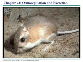
Freshwater animals show adaptations that reduce water uptake and conserve solutes • Desert and marine animals face desiccating environments that can quickly deplete body water • Excretion gets rid of nitrogenous metabolites and other waste products
Concept 44.1: Osmoregulation balances the uptake and loss of water and solutes • Osmoregulation is based largely on controlled movement of solutes between internal fluids and the external environment
Osmosis and Osmolarity • Cells require a balance between osmotic gain and loss of water • Osmolarity, the solute concentration of a solution, determines the movement of water across a selectively permeable membrane • If two solutions are isoosmotic, the movement of water is equal in both directions • If two solutions differ in osmolarity, the net flow of water is from the hypoosmotic to the hyperosmotic solution
Fig. 44-2 • Selectively permeable • membrane • Solutes • Net water flow • Water • Hypoosmotic side • Hyperosmotic side
Osmotic Challenges • Osmoconformers, consisting only of some marine animals, are isoosmotic (same osmotic concentration) with their surroundings and do not regulate their osmolarity • Osmoregulators expend energy as ATP to control water uptake and loss in a hyperosmotic or hypoosmotic environment
Marine Animals • Most marine invertebrates are osmoconformers • Most marine vertebrates and some invertebrates are osmoregulators • Marine bony fishes are hypoosmotic to sea water • They lose water by osmosis and gain salt by diffusion and from food • They balance water loss by drinking seawater and excreting salts
Fig. 44-4a • Excretion • of salt ions • from gills • Osmotic water • loss through gills • and other parts • of body surface • Gain of water and • salt ions from food • Gain of water • and salt ions from • drinking seawater • Excretion of salt ions and • small amounts of water in • scanty urine from kidneys • (a) Osmoregulation in a saltwater fish
Freshwater Animals • Freshwater animals constantly take in water by osmosis from their hypoosmotic environment • They lose salts by diffusion and maintain water balance by excreting large amounts of dilute urine • Salts lost by diffusion are replaced in foods and by uptake across the gills
Fig. 44-4b • Osmotic water • gain through gills • and other parts • of body surface • Uptake of water and • some ions in food • Uptake • of salt ions • by gills • Excretion of large • amounts of water in • dilute urine from kidneys • (b) Osmoregulation in a freshwater fish
Animals That Live in Temporary Waters • Some aquatic invertebrates in temporary ponds lose almost all their body water and survive in a dormant state
Fig. 44-5 • 100 µm • 100 µm • (b) Dehydrated • tardigrade • (a) Hydrated tardigrade
Fresh water protists have contractile vacuoles • https://www.youtube.com/watch?v=9Ynm5ZOW59Q • http://www.gettyimages.com/detail/video/amoeba-proteus-protozoan-stock-video-footage/618601067
Land Animals • Land animals manage water budgets by drinking and eating moist foods and using metabolic water (water produced in cell respiration) • Desert animals get major water savings from simple anatomical features and behaviors such as a nocturnal life style
Fig. 44-6 • Water • balance in a • kangaroo rat • (2 mL/day) • Water • balance in • a human • (2,500 mL/day) • Ingested • in food (0.2) • Ingested • in food (750) • Ingested • in liquid (1,500) • Water • gain • (mL) • Derived from • metabolism (250) • Derived from • metabolism (1.8) • Feces (0.09) • Feces (100) • Water • loss • (mL) • Urine • (1,500) • Urine • (0.45) • Evaporation (1.46) • Evaporation (900)
Energetics of Osmoregulation • Osmoregulators must expend energy to maintain osmotic gradients
Transport Epithelia in Osmoregulation • Animals regulate the composition of body fluid that bathes their cells • Transport epithelia are specialized epithelial cells that regulate solute movement • They are essential components of osmotic regulation and metabolic waste disposal • They are arranged in complex tubular networks • An example is in salt glands of marine birds, which remove excess sodium chloride from the blood
Fig. 44-7 • EXPERIMENT • Nasal salt • gland • Ducts • Nostril • with salt • secretions
Fig. 44-8 • Vein • Artery • Secretory • tubule • Secretory • cell • Salt gland • Capillary • Secretory tubule • Transport • epithelium • NaCl • NaCl • Direction of • salt movement • Central duct • Blood • flow • Salt secretion • (b) • (a)
Concept 44.2: An animal’s nitrogenous wastes reflect its phylogeny and habitat • The type and quantity of an animal’s waste products may greatly affect its water balance • Among the most important wastes are nitrogenous breakdown products of proteins and nucleic acids • Some animals convert toxic ammonia (NH3) to less toxic compounds prior to excretion
Fig. 44-9 • Proteins • Nucleic acids • Amino • acids • Nitrogenous • bases • Amino groups • Most aquatic • animals, including • most bony fishes • Mammals, most • amphibians, sharks, • some bony fishes • Many reptiles • (including birds), • insects, land snails • Ammonia • Uric acid • Urea
Forms of Nitrogenous Wastes • Different animals excrete nitrogenous wastes in different forms: ammonia, urea, or uric acid
Ammonia • Animals that excrete nitrogenous wastes as ammonia need lots of water • They release ammonia across the whole body surface or through gills
Urea • The liver of mammals and most adult amphibians converts ammonia to less toxic urea • The circulatory system carries urea to the kidneys, where it is excreted • Conversion of ammonia to urea is energetically expensive; excretion of urea requires less water than ammonia
Uric Acid • Insects, land snails, and many reptiles, including birds, mainly excrete uric acid • Uric acid is largely insoluble in water and can be secreted as a paste with little water loss • Uric acid is more energetically expensive to produce than urea
The Influence of Evolution and Environment on Nitrogenous Wastes • The kinds of nitrogenous wastes excreted depend on an animal’s evolutionary history and habitat • The amount of nitrogenous waste is coupled to the animal’s energy budget
Concept 44.3: Diverse excretory systems are variations on a tubular theme • Excretory systems regulate solute movement between internal fluids and the external environment
Excretory Processes • Most excretory systems produce urine by refining a filtrate derived from body fluids • Key functions of most excretory systems: • Filtration: pressure-filtering of body fluids • Reabsorption: reclaiming valuable solutes • Secretion: adding toxins and other solutes from the body fluids to the filtrate • Excretion: removing the filtrate from the system
Fig. 44-10 • Filtration • Capillary • Filtrate • Excretory • tubule • Reabsorption • Secretion • Urine • Excretion
Survey of Excretory Systems • Systems that perform basic excretory functions vary widely among animal groups • They usually involve a complex network of tubules
Fig. 44-11 • Nucleus • of cap cell • Cilia • Flame • bulb • Interstitial • fluid flow • Opening in • body wall • Tubule • Tubules of • protonephridia • Tubule cell
Fig. 44-12 • Coelom • Capillary • network • Components of • a metanephridium: • Internal opening • Collecting tubule • Bladder • External opening
Fig. 44-13 • Digestive tract • Rectum • Hindgut • Intestine • Midgut • (stomach) • Malpighian • tubules • Salt, water, and • nitrogenous • wastes • Feces and urine • Rectum • Reabsorption • HEMOLYMPH
Kidneys • Kidneys, the excretory organs of vertebrates, function in both excretion and osmoregulation
Structure of the Mammalian Excretory System • The mammalian excretory system centers on paired kidneys, which are also the principal site of water balance and salt regulation • Each kidney is supplied with blood by a renal artery and drained by a renal vein • Urine exits each kidney through a duct called the ureter • Both ureters drain into a common urinary bladder, and urine is expelled through a urethra
Fig. 44-14a • Posterior • vena cava • Renal artery • and vein • Kidney • Aorta • Ureter • Urinary • bladder • Urethra • (a) Excretory organs and major • associated blood vessels
Fig. 44-14ab • Renal • medulla • Posterior • vena cava • Renal • cortex • Renal artery • and vein • Kidney • Renal • pelvis • Aorta • Ureter • Urinary • bladder • Ureter • Urethra • Section of kidney • from a rat • (a) Excretory organs and major • associated blood vessels • (b) Kidney structure • 4 mm
The nephron, the functional unit of the vertebrate kidney, consists of a single long tubule and a ball of capillaries called the glomerulus • Bowman’s capsule surrounds and receives filtrate from the glomerulus
Fig. 44-14c • Juxtamedullary • nephron • Cortical • nephron • Renal • cortex • Collecting • duct • Renal • medulla • To • renal • pelvis • (c) Nephron types
Fig. 44-14d • Glomerulus • Afferent arteriole • from renal artery • Bowman’s capsule • 10 µm • SEM • Proximal tubule • Peritubular capillaries • Efferent • arteriole from • glomerulus • Distal • tubule • Branch of • renal vein • Collecting • duct • Descending • limb • Loop of • Henle • Ascending • limb • Vasa • recta • (d) Filtrate and blood flow
Filtration of the Blood • Filtration occurs as blood pressure forces fluid from the blood in the glomerulus into the lumen of Bowman’s capsule • Filtration of small molecules is nonselective • The filtrate contains salts, glucose, amino acids, vitamins, nitrogenous wastes, and other small molecules
Pathway of the Filtrate • From Bowman’s capsule, the filtrate passes through three regions of the nephron: the proximal tubule, the loop of Henle, and the distal tubule • Fluid from several nephrons flows into a collecting duct, all of which lead to the renal pelvis,which is drained by the ureter
Blood Vessels Associated with the Nephrons • Each nephron is supplied with blood by an afferent (going to) arteriole, a branch of the renal artery that divides into the capillaries • The capillaries converge as they leave the glomerulus, forming an efferent (going from) arteriole • The vessels divide again, forming capillaries, which surround the proximal and distal tubules
There are specific capillaries that serve the loop of Henle • These capillaries and the loop of Henle function as a countercurrent system
Concept 44.4: The nephron is organized for stepwise processing of blood filtrate • The mammalian kidney conserves water by producing urine that is much more concentrated than body fluids
From Blood Filtrate to Urine: A Closer Look Proximal Tubule • Reabsorption of ions, water, and nutrients takes place in the proximal tubule • Molecules are transported actively and passively from the filtrate into the interstitial fluid and then capillaries • Some toxic materials are secreted into the filtrate • The filtrate volume decreases Animation: Bowman’s Capsule and Proximal Tubule
Descending Limb of the Loop of Henle • Reabsorption of water continues through channels formed by aquaporin proteins • Movement is driven by the high osmolarity of the interstitial fluid, which is hyperosmotic to the filtrate • The filtrate becomes increasingly concentrated
Ascending Limb of the Loop of Henle • In the ascending limb of the loop of Henle, salt but not water is able to diffuse from the tubule into the interstitial fluid • The filtrate becomes increasingly dilute