Download
1 / 34
340 likes | 343 Views
This text discusses the major functions of the digestive system, the necessity of digestion, the organs involved, the absorption of nutrients, and common disorders and diseases. It also explains the types of digestion, the six processes involved in digestion, and the structure of the alimentary canal and accessory digestive organs. The text is written in English.

E N D
Objectives: • List and describe the major functions of the digestive system. • Describe why digestion of food is necessary and compare mechanical and chemical digestion • Identify the major digestive organs, the digestive accessory glands, and their functions • Describe the absorption of food in the small intestine and the absorption of water in the large intestine • List and describe disorders and diseases of the digestive system.
Function • Digestion • Breakdown of ingested food • Absorption of nutrients into the blood • Once the nutrients are absorbed by the digestive system they are transported by the blood to the tissues for metabolism. • Production of cellular energy (ATP) • Constructive and degradative cellular activities
Types of Digestion • Digestion is a catabolic process in which large complex molecules (carbohydrates, lipids, proteins, nucleic acids) are broken down into simpler monomers (monosaccharides, glycerol and fatty acids, amino acids, and nucleotides) which can be absorbed by the body. • There are two forms of digestion: a. mechanical: In mechanical there is no chemical change in the food. The food is simply broken down into smaller pieces and mixed with digestive juices secreted in the body. Ex. Mastication (chewing) b. chemical: In chemical digestion the is a chemical change in the food. The polymers are broken down into monomers commonly by hydrolysis reactions carried out by enzymes contained within the digestive juices.
Six Processes of Digestion • Ingestion – getting food into the mouth • Propulsion – moving foods from one region of the digestive system to another • Mechanical digestion • Mixing of food in the mouth by the tongue • Churning of food in the stomach • Segmentation in the small intestine • Chemical Digestion • Enzymes break down food molecules into their building blocks • Each major food group uses different enzymes • Carbohydrates are broken to simple sugars • Proteins are broken to amino acids • Fats are broken to fatty acids and alcohols • Absorption • End products of digestion are absorbed in the blood or lymph • Food must enter mucosal cells and then into blood or lymph capillaries • Defecation • Elimination of indigestible substances as feces
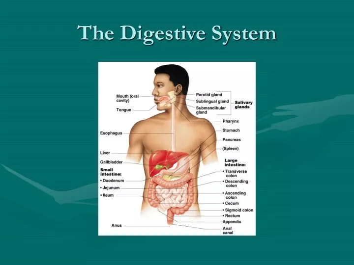
Divisions of Digestive System Organs • Two main groups • Alimentary canal – continuous coiled hollow tube that runs from the mouth to the anus • Accessory digestive organs- secrete digestive juices by ducts (exocrine glands) into the alimentary canal.
Organs of the Alimentary Canal • Mouth • Pharynx • Esophagus • Stomach • Small intestine • Large intestine • Anus
Accessory Digestive Organs • Salivary glands • Teeth • Pancreas • Liver • Gall Bladder
Mouth Oral Cavity (Alimentary Canal) • Mastication (chewing) of food • Mixing masticated food with saliva • Initiation of swallowing by the tongue • Allowing for the sense of taste
Salivary Glands (Accessory Organs) Salivary Glands: • Saliva-producing glands • Parotid glands – located anterior to ears • Submandibular glands • Sublingual glands Saliva: • Mixture of mucus and serous fluids • Helps to form a food bolus • Contains salivary amylase to begin starch digestion • Dissolves chemicals so they can be tasted
Teeth (Accessory Organs) • The role is to masticate (chew) food • Humans have two sets of teeth • Deciduous (baby or milk) teeth • 20 teeth are fully formed by age two • Permanent teeth • Replace deciduous teeth beginning between the ages of 6 to 12 • A full set is 32 teeth, but some people do not have wisdom teeth • Types of teeth: Incisors - cutting Canines - tearing Premolars – shearing, shredding Molars - grinding
Tooth Structure • Crown – exposed part • Outer enamel • Dentin • Pulp cavity • Neck • Region in contact with the gum • Connects crown to root • Root • Periodontal membrane attached to the bone • Root canal carrying blood vessels and nerves
Pharynx (Alimentary Canal) • Serves as a passageway for air and food • Food is propelled to the esophagus by two muscle layers • Longitudinal inner layer • Circular outer layer • Food movement is by alternating contractions of the muscle layers (peristalsis)
Esophagus (Alimentary Canal) • Runs from pharynx to stomach through the diaphragm • Conducts food by peristalsis (slow rhythmic squeezing) • Passageway for food only (respiratory system branches off after the pharynx)
Alimentary Canal Organ Structure and Tissue Arrangement • Mucosa • Innermost layer • Moist membrane • Surface epithelium • Small amount of connective tissue (lamina propria) • Small smooth muscle layer • Submucosa • Just beneath the mucosa • Soft connective tissue with blood vessels, nerve endings, and lymphatics • Muscularis externa – smooth muscle • Inner circular layer • Outer longitudinal layer • Serosa • Outermost layer – visceral peritoneum • Layer of serous fluid-producing cells
Stomach (Alimentary Canal) • Located on the left side of the abdominal cavity • Food enters at the cardioesophageal sphincter • Regions of the stomach • Cardiac region – near the heart • Fundus • Body • Pylorus – funnel-shaped terminal end • Food empties into the small intestine at the pyloric sphincter • Rugae – internal folds of the mucosa • External regions • Lesser curvature • Greater curvature
Stomach (Alimentary Canal) • Acts as a storage tank for food • Site of food breakdown • Chemical breakdown of protein begins • Delivers chyme (processed food) to the small intestine
Stomach: Mucosal Layer • Simple columnar epithelium • Mucous neck cells (goblet cells) – produce a sticky alkaline mucus • Gastric glands – secrete gastric juice • Chief cells – produce protein-digesting enzymes (pepsinogens) • Parietal cells – produce hydrochloric acid • Endocrine cells – produce gastrin • Gastric pits formed by folded mucosa • Glands and specialized cells are in the gastric gland region
Small Intestine (Alimentary Canal) • The body’s major digestive organ all digestion of food is completed in this organ • Site of nutrient absorption into the blood • Muscular tube extending form the pyloric sphincter to the ileocecal valve • Suspended from the posterior abdominal wall by the mesentery • Duodenum (25cm = 10 inches) “12 finger widths long” • Attached to the stomach • Curves around the head of the pancreas • Where bile and pancreatic juices enter the alimentary canal • Jejunum (2.5m = 8 feet) “empty” • Attaches anteriorly to the duodenum • Ileum (3.6m = 12 feet) “twisted” • Extends from jejunum to large intestine
Small Intestine Internal Structure • Villi are small fingerlike structures formed by the mucosa • Give the small intestine more surface area for absorption • Fold in the intestine are called circular folds or plicae circulares • Deep folds of the mucosa and submucosa • Do not disappear when filled with food • The submucosa has Peyer’s patches (collections of lymphatic tissue)
Villi Internal Structure and Function • Absorptive cells are found on the surface epithelium which are simple columnar microvilliated epithelium • Blood capillaries are below the surface epithelium and this is where monosaccharides, amino acids, and nucleic acids enter into the blood stream and are taken to the liver for processing • Lacteals (specialized lymphatic capillaries) where lipids are absorbed and eventually re-enter the blood stream to be taken to the liver for processing.
Pancreas (Accessory Organ) • Produces a wide spectrum of digestive enzymes that break down all categories of food • Enzymes are secreted into the duodenum • Alkaline fluid introduced with enzymes neutralizes acidic chyme • Endocrine products of pancreas • Insulin • Glucagon
Liver and Gall Bladder (Accessory Organs) • Largest gland in the body • Located on the right side of the body under the diaphragm • Consists of four lobes suspended from the diaphragm and abdominal wall by the falciform ligament • Connected to the gall bladder via the common hepatic duct • Produced by cells in the liver • Composition • Bile salts • Bile pigment (mostly bilirubin from the breakdown of hemoglobin) • Cholesterol • Phospholipids • Electrolytes • Sac found in hollow fossa of liver • Stores bile from the liver by way of the cystic duct • Bile is introduced into the duodenum in the presence of fatty food • Gallstones can cause blockages
Large Intestine (Alimentary Canal) • Larger in diameter, but shorter than the small intestine • Frames the internal abdomen • Cecum – saclike first part of the large intestine • Appendix • Accumulation of lymphatic tissue that sometimes becomes inflamed (appendicitis) • Hangs from the cecum • Colon • Ascending • Transverse • Descending • S-shaped sigmoidal • Rectum • Anus – external body opening
Functions of Large Intestine • Absorption of water • Eliminates indigestible food from the body as feces • Does not participate in digestion or absorption of digested food • Goblet cells produce mucus to act as a lubricant • Site of production of Vitamin K by symbiotic bacteria which live off the remains of food that have not been digested or absorbed in the small intestine. These bacteria produce over 50% of fecal matter.
Nutrition • Nutrient – substance used by the body for growth, maintenance, and repair. Macronutrients are those which are required in large amounts. Micronutrients required in smaller amounts. • Categories of nutrients • Carbohydrates ( macro) • Lipids (macro) • Proteins (macro) • Vitamins (micro) • Mineral (micro) • Water • A lack of the proper nutrients or an imbalance in the correct amounts of each is called malnutrition. Even though a person is obese they often suffer from malnutrition!
Source of Nutrients • Carbohydrates • Most are derived from plants • Exceptions: lactose from milk and small amounts of glycogens from meats • Lipids • Saturated fats from animal products • Unsaturated fats from nuts, seeds, and vegetable oils • Cholesterol from egg yolk, meats, and milk products • Proteins • Complete proteins – contain all essential amino acids • Most are from animal products • Legumes and beans also have proteins, but are incomplete • Vitamins • Most vitamins are used as cofactors and act with enzymes many are produced by plants • Found in all major food groups • Minerals • Play many roles in the body • Most mineral-rich foods are vegetables, legumes, milk, and some meats
Diseases and Disorders of the Digestive System • Heartburn (Acid Reflux) This is due to acid from the stomach entering into the esophagus which results in a burning sensation. In chronic severe cases this can lead to damage, ulceration, scarring, and possibly cancer of the esophagus if not treated.
Diseases and Disorders of the Digestive System • Ulcers: Ulcers occur when the lining of the stomach or the duodenum becomes weakened and exposed to the effects of digestive enzymes and stomach acid. It eventually will digest a hole through the mucosa and may cause severe bleeding if a blood vessel of the stomach is involved. It is now known that ulcers are closely associated with infection by a bacteria called Helicobacter pylori.
Diseases and Disorders of the Digestive System • Crohn’s Disease: Crohn's Disease is an inflammatory disease of the bowel. It can cause fever, pain, diarrhoea and significant loss of weight. Crohn's Disease can affect any part of the bowel, but most typically affects the lower end of the small intestine, where it joins the large intestine. The intestinal wall becomes thick and inflamed, producing ulcers and fissures, and can in addition cause abnormal passageways to form between adjacent portions of the intestine. The intestinal space becomes so narrow that the passage of food can become obstructed.
Diseases and Disorders of the Digestive System • Colon Cancer: Cancer starts in the inner layer and can grow through some or all of the other layers. Knowing a little about these layers is helpful because the stage (extent of spread) of a cancer depends to a great degree on which of these layers it affects. • Cancer that starts in the different areas may cause different symptoms. Colon and rectum cancers probably develop slowly over a period of several years. We now know that most of these cancers begin as a polyp--a growth of tissue into the center of the colon or rectum. Polyps are also known as adenomas. Removing the polyp early may prevent it from becoming cancer. • Over 95% of colon and rectal cancers are adenocarcinomas. These are cancers of the cells that line the inside of the colon and rectum. There are some other, more rare, types of tumors of the colon and rectum, but the facts given here refer only to adenocarcinomas. • Colon and rectal cancer have many features in common and are often referred to together as colorectal cancer.