Download
1 / 120
1.2k likes | 1.21k Views
Ultrasound of the Abdomen Part II lecture 12: Retroperitoneum Part B HHHoldorf. Outline. The Abdomen - Overview Abscess Biloma Ascites Lymphocele Urinoma. Pseudomyxoma Peritonei Hematoma Lymphoma Thorax (non-Cardiac Chest) Baker’s cyst Rectus Sheath Hematoma Abdominal Wall neoplasm

E N D
Ultrasound of the Abdomen Part IIlecture 12: Retroperitoneum Part BHHHoldorf
Outline • The Abdomen - Overview • Abscess • Biloma • Ascites • Lymphocele • Urinoma • Pseudomyxoma Peritonei • Hematoma • Lymphoma • Thorax (non-Cardiac Chest) • Baker’s cyst • Rectus Sheath Hematoma • Abdominal Wall neoplasm • Hernias/Inguinal Hernia
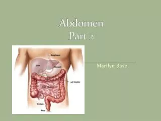
Superiorly….Diaphragm • Inferiorly….Pelvis • Anteriorly…Abdominal Wall muscle • Posteriorly…Vertebral column, ribs, Iliac fossa. Boundaries of The abdominal cavity
Xiphoid process • Palpated where the costal margins meet at the infra-sternal angle • Costal margins • Curved lower margin of the thoracic wall • Iliac crest • Palpated and ends in front at the anterior iliac spine and behind at the posterior iliac spine • Pubic symphysis • Cartilaginous joint that lies in the midline between bodies of the pubic bones • Linea albea (white Line) • Midline fibrous band that extends from the pubic symphysis to the Xiphoid process • Umbilicus • Remnant of the fetal umbilical cord Surface landmarks of the anterior abdominal wall
From outermost layer working in: Skin- epidermis is highly echogenic Superficial fascia Subcutaneous fat- relatively anechoic. Muscle layers Transversalis fascia Extraperitoneal fat Anatomy of Anterior Abdominal Wall
Rectus sheath- Fibrotic band which extends from the Xiphoid process to the symphysis pubis. Linea Alba- The rectus sheath joins in the midline to form linea alba, separating the rectus muscles in the midline. Linea semilunaris- A curved tendinous line placed one on either side of the rectus Abdominis. Arcuate line- Located midway between the umbilicus and symphysis pubis. At this level the post caudal portion of the rectus sheath ends and the joining of all three muscles pass in front of the rectus muscle. Anterior abdominal muscles cont.
Psoas Muscle • Medial and posterior to kidneys • Lateral to spine • Quadratus Lumborum • Adjacent to the iliac crest • Depending on the level; posterior to kidneys, colon, psoas m. • Tapers cephalically (Sag.) • Iliacus Muscle-within the iliac space • iliopsoas Muscle-A convergence of the iliacus and psoas muscle. Posterior Abdominal Wall Muscles
A large muscle • Forms partition between thoracic and abdominal cavities • Dome shaped • Moves with respiration- Due to large effusion or bronchial Cancer the diaphragm might not move with respiration and become flatten or Convex • Normal US- concave, echogenic • Appearance can change due to hernia, pleural effusion, COPD (Chronic obstructive pulmonary disease) Diaphragm
The Aorta, IVC, esophagus pass through individual openings called Hiatuses in the diaphragm. • Crura: Are elongated muscular bands that arise from the lumbar vertebrae and insert into the diaphragm and connect the diaphragm and the spinal column. On the Us Crus appears as low level echoes. • The right crus- Post. to IVC • The left crus- Ant. to Aorta, proximal to the celiac trunk • Attachments: • Attached to Xiphoid process in front • Attached to costal margins at sides • Rt. & Lt. parts insert into the central tendon. Diaphragm cont.
How does the Diaphragm move with respiration? How does this affect lower extremity Venous return?
The sonographic appearance of an abscess is quite variable. Typically, an abscess is a complex mass (solid or cystic). Debris, septations and gas can be seen within the abscess. The boarders of an abscess are typically irregular. • Gas within the abscess may produce a reverberation (comet-tail) artifact. • Abscesses typically demonstrate posterior enhancement depending on the cystic component of the abscess. • The most reliable findings in patients with abscesses are: Presence of fever Increased white blood cell count ABSCESS
Variable appearance on US: • Generally hypo to anechoic • some may contain internal debris. • Usually has acoustic enhancement. • Presence of gas produces bright echogenic reflections and dirty shadow Abscess:
Bilomas are extrahepatic collections of extravasated bile. They are caused by abdominal trauma, gallbladder disease, or biliary surgery. Bilomas are predominantly cystic masses located in the right upper quadrant. Biloma
Extrahepatic collection of extravagated bile. • Causes: trauma, GB disease, biliary surgery. • Ultrasound appearance: • Anechoic mass with sharp borders & good through transmission. • May contain some debris. • Located in RUQ or mid abdomen. Biloma:
Ascites is the excessive accumulation of serous fluid in the peritoneal cavity. The mechanisms that produce Ascites are complex and incompletely understood. • Two mechanisms that produce Ascites are: • Low serum osmotic pressure (protein Loss) • High portal venous pressure • Causes of Ascites include: • Cirrhosis (most common cause) • Hypoalbuminemia (decreased protein) • Budd-Chiari Syndrome • Heart failure • Cancer • Nephrotic syndrome (protein loss) Ascites
Hypoalbuminemia (low protein) can be the result of liver failure, nephritic syndrome or malnutrition. • Transjugular intrahepatic portal systemic shunting (TIPS) can successfully treat Ascites by lowering portal pressure. • Ascites is commonly found at the • Inferior aspect of the right lobe of the liver • Morison’s pouch • Pelvic cul de sac • Paracolic gutters
Gallbladder wall thickening is frequently seen with Ascites Benign Ascites is indicated by freely floating bowel. With malignant Ascites, the bowel loops are tethered or matted to the posterior abdominal wall surrounded by complex or loculated fluid collections.
Pleural effusion- fluid collection superior to the diaphragm • Ascites- Excessive accumulation of serous free fluid surrounding or interposed between organs. Collects Inferior to the diaphragm. Over 90% are due to Cirrhosis, Neoplasm, or CHF. Other causes: Hypoalbuminemia, endocrine disease. Pancreatic Ascites may develop due to chronic pancreatitis or Pancreatic pseudocyst.
Transudative ascites is defined as having less than 3 g of protein per 100 ml of fluid. It is, as its name would suggest, a transudate* - a result of raised hydrostatic pressure forcing fluid out of blood vessels. Causes include: cardiac failure. *A filtrate of blood Ascites: Transudative vs. Exudative
Exudate ascites is defined as ascites with a protein content of greater than 3g protein per 100ml of fluid. Possible causes of exudate ascites include: malignant disease. pyogenic infection. tuberculosis.
Transudative • US: Anechoic region in peritoneal cavity. • Collects in the most dependent area of the abdomen • First seen in the Morrison’s pouch or pouch ofDouglas. • then seen in Paracolic gutters. • Freely mobile, bowel loop can be seen floating within ascites. Ascites cont.
Exudative • Due to inflammation and malignancy. • US: May have Septations, Echogenic debris, loculation of fluid, matted bowel loops. • Loculated ascites: is no compressible, does not move with change of position. • Differential Diagnosis: Abscess, cystic Neoplasm, hematoma, lymphocele. Ascites cont.
Urine ascites: results from renal transplant, pt. on chronic hemodialysis. Bloody Ascites: caused by ruptured Aorta, ruptured ectopic pregnancy, etc. Ascites
Lymphoceles are complications of Renal transplantation Gynecologic surgery Vascular Surgery Urological Surgery Caused by leakage of lymph from a renal allograft (transplant) or by a surgical disruption of the lymphatic channels. Differential diagnosis includes any fluid collection such as loculated Ascites, Urinoma, hematoma, or abscess. The presence of internal echoes within the collection is more consistent with an abscess or hematoma than with a Lymphocele. Lymphocele
A lymph–filled cystic mass with no epithelial lining. • Causes: • Complication of surgery ( involving-vascular, urological, gynecological, renal.) • Leak by surgical disruption of the lymph. • US: round,/elliptical, anechoic, septations seen frequently, w/ good through transmission Lymphocele
An Urinoma is a collection of urine which is located outside of the kidney or bladder. Urinomas are caused by renal trauma, renal surgery, or from an obstructing lesion. Most commonly associated with Renal transplantation. Posterior urethral valve obstruction May accumulate directly after a renal transplant due to an anastomotic leak of the ureter. sonographic appearance is similar to a Lymphocele. Urinoma
A collection of urine, located outside the kidney or bladder due to an obstructing lesion. Causes : Renal trauma, renal surgery, renal transplant. US: elliptical anechoic mass located in perinephric space. Differential Diagnosis: Lymphocele. Urinoma:
The filling of the peritoneal cavity with mucinous material and gelatinous Ascites. Tumor implants are found on the peritoneal surfaces. Bowel loops will be matted to the posterior abdominal wall. Caused by metastasis or rupture of a mucinous cystadenocarcinoma of the ovary or mucinous tumor of the appendix. This is referred to as malignant Ascites. Pseudomyxoma Peritonei
A hematoma is a collection of blood which is usually confined to an organ, tissue or space. The ultrasound appearance of hematomas is variable and depends on the age of the collection. Fibrin invasion causes hematomas to appear hyperechoic. Gradual hemolysis eventually creates an anechoic appearance. Organization of clot or fragmentation of the clot will produce irregular echoes. Calcifications are often associated with longstanding hematomas. A decrease in the hematocrit level indicates the presence of a hematoma. Hematocrit is the volume of red blood cells fond in 100 ml of blood. Blood spillage outside the circulatory system will result in a decreased hematocrit Hematomas