Download
1 / 17
210 likes | 281 Views
Histology of digestive system stomach-fundus & pylorus. INTRODUCTION. BASIC PATTERN: I) Mucosa - Epithelium - Lamina propria - Muscularis Mucosae II) Submucosa III) Muscularis Externa IV) Serous layer / Adventitial layer. STOMACH.

E N D
INTRODUCTION • BASIC PATTERN: I) Mucosa - Epithelium - Lamina propria - Muscularis Mucosae II) Submucosa III) Muscularis Externa IV) Serous layer / Adventitial layer
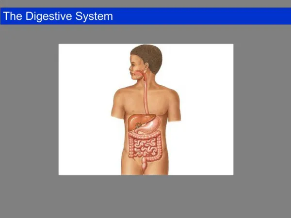
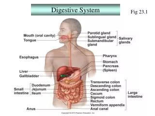
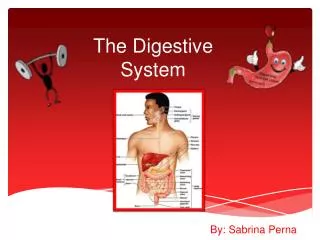
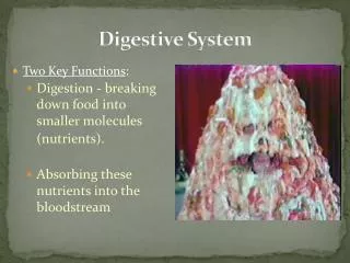
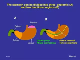
STOMACH • Stomach is a dilated segment of the digestive tract, that digest food and secrets hormone • There are three histological regions: • Cardia • Fundus and body • Pylorus • The fundus and body are identical in microscopic structure • The mucosa and submucosa of the undistended stomach lie in longitudinally directed folds known as rugae • When the stomach is filled with food, folds flatten out
Cardiac region of the stomach Mucosa: • Epithelial lining at the cardio-oesophageal junction changes from stratified sq. to simple columnar epithelium • Presence of Mucous surface cells and cardiac glands Submucosa: Consists of Meissner’s plexus and blood vessels
Muscularis externa: Inner – Oblique Middle – circular Outer – longitudinal Myenteric plexus between muscle layers Serosa – Simple squamous epithelium resting on a thin layer of connective tissue
Stomach - fundus • Mucosa: - Lining Epithelium – Simple columnar epithelium that invaginates to various extents into the lamina propria, forming gastric pits. - These cells are involved in mucus secretion. The mucus protects the epithelial lining from damage due to the presence of acid in the stomach.
Stomach – fundus • Lamina propria: - Small tubular fundic/gastric glands are present. - Types of cells: i) Mucous neck cells – Located just below gastric pit. - Columnar in shape - Contain mucinogen granules in apical cytoplasm, while nuclei are situated basally. - Produces soluble mucus
Parietal or oxyntic cells: - They are large, ovoid or polyhedral cells with a large central nucleus. - More numerous in the upper half of the gland than in the lower half - Secretes HCL and intrinsic factor. Intrinsic factor combines with vitamin B12 to form a complex necessary for erythrocytes formation.
Chief or zymogenic cells: - Located in the lower 1/3rd of gastric glands. - Contain rough endoplasmic reticulum near the base, secretory granules near their apex and a small golgi apparatus. - Secrete pepsinogen which is converted into pepsin in an acid environment.
Enteroendocrine cells: - Located in the basal portion of gastric glands - Secretes serotonin, histamine and gastrin. These are endocrine cells which release their products into the blood vessels. MUSCULARIS MUCOSAE: - It consists of two thin layer of smooth muscles. i.e., Outer longitudinal and inner circular
SUBMUCOSA: - Consists of blood vessels, lymphatic vessels and Meissner’s plexus. MUSCULARIS EXTERNA: Inner: Oblique Middle: circular Outer: Longitudinal SEROSA: Outermost layers of the stomach which consists of loose connective tissue covered by mesothelium
STOMACH - PYLORUS • MUCOSA: - Epithelium: Simple columnar as in fundic part - Pyloric glands occupy the lamina propria - Gastric pits are deeper - Glands are short,tortuous and branched - Produce mucus and gastrin - Muscularis mucosa and Submucosa are similar to fundic part
MUSCULARIS EXTERNA: Inner: Oblique Middle: circular Outer: Longitudinal Similar to Fundic part, but the circular fibres are much thickened to form pyloric sphincter
CLINICAL ASPECTS • GASTRO- ESOPHAGEAL REFLUX DISORDER: A condition which is characterised by the compromised state of the lower oesophageal sphincter, which leads to reflux of food and acid into oesophagus.