Download
1 / 50
500 likes | 530 Views
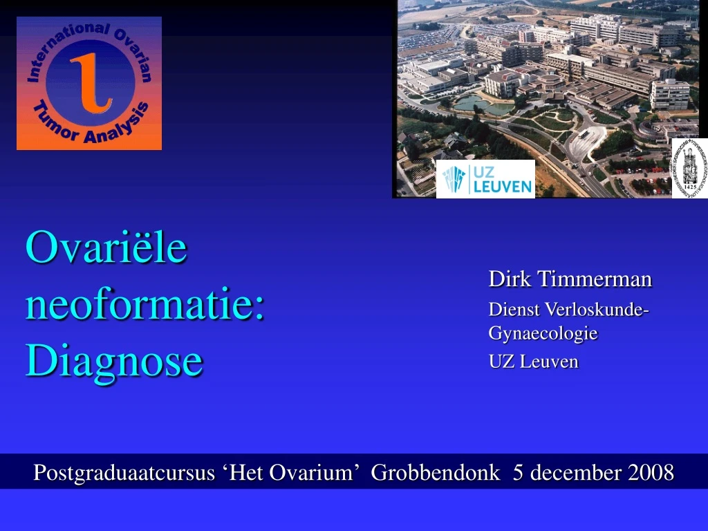
Ovariële neoformatie : Diagnose. Dirk Timmerman Dienst Verloskunde-Gynaecologie UZ Leuven. Postgraduaatcursus ‘ Het Ovarium ’ Grobbendonk 5 december 2008. Meta-Analysis (n = 1 545 ) Invasive Ovarian Cancer Stage I. 5 independent variables for decreased disease-free survival :

E N D
Ovariële neoformatie: Diagnose Dirk Timmerman Dienst Verloskunde-Gynaecologie UZ Leuven Postgraduaatcursus ‘HetOvarium’ Grobbendonk 5 december 2008
Meta-Analysis (n = 1545)Invasive Ovarian Cancer Stage I • 5 independent variables for decreased disease-free survival: differentiation, rupture before and during surgery, FIGO 1973, and age. • Laparoscopic removal should be restricted to benign ovarian cysts (Vergote et al. Lancet 2001;357:176-82)
1994: confusing… • Clinical examination • Morphologic scoring systems • Risk of malignancy index (RMI) • Colour Doppler Imaging: PI or RI? • Serum CA 125?
Morphologic Scoring Systems:prospective comparison (n=330) (Ferrazzi E, et al. Ultrasound Obstet Gynecol 1997;10:192-7)
Risk of Malignancy Index RMI = U x M x CA125 • U = Ultrasound score • 0 : no features (multiloc. cysts, solid areas, metastases, ascites) • 1 : one feature • 2 : two or more features • M = Menopausal status • 1 : premenopausal • 3 : postmenopausal (Jacobs, 1990)
Benign Malignant
Colour score • 1 : no colour • 2 : minimal colour • 3 : rather strong • 4 : very strong (Leuven) Endometrioid ovarian cancer
Development set (1994-1996) 173 patients benign : 124 malignant : 49 Prospective set (1996-1999) 257 patients benign : 170 malignant : 87 Data Description Patient data collected at University Hospitals Leuven
Logistic regression model Papillarities >3mm (0,1) Color score (1,2,3,4) ~Probability of cancer Menopause (0,1) CA 125 (1 - 31 090)
Neural Network Model Input layer of source nodes Layer of hidden neurons Output neuron Papillarities >3mm (0,1) Smooth wall (0,1) Unilocular (0,1) ~Probability of cancer Ascites (0,1) Bilateral (0,1) Menopause (0,1) CA 125 (1 - 31 090) (Ultrasound Obstet Gynecol 1999;13:17-25)
A, B (= 5,000- 10,000 TVS) D, E, F (= 200- 300 TVS) C (= 1,000 TVS) Subjective assessment (n=300) Sensitivity False pos. rate (Ultrasound Obstet Gynecol. 1999)
‘Ground glass’ Anechoic Low echogenicity Hemorrhagic Mixed Mixed (old blood-fluid or fat-fluid) Mixed (e.g. abscess) (IOTA)
Colour distribution • simple cyst, follicle: no or minimal (wall) • preovulatory follicle: more flow (wall) • corpus luteum (cyst): strong,surrounding • endometrioma : minimal,localised • benign tumour: minimal or rather strong (septa+wall) • tubo-ovarian abscess: strong (septa+wall) • ovarian cancer: strong (septa+wall)
Role of colour Doppler (CDE) in the diagnosis of endometrioma (N= 58) B-mode Typical Atypical CDE evaluation Non endometrioma Endometrioma Non endometrioma (Guerriero S, Ajossa S et al (Cagliari) Human Reprod 1998; 13: 1691-5)
Criticism • Retrospective evaluation of new models • Generalisability? (‘over-fitting’)
New developments • New US techniques (3D, i.v. contrast…) • Multicentre prospective studies • New models with built-in expertise • New biochemical markers (proteomics)
Dirk Timmerman, Leuven Lil Valentin, Malmö Antonia Testa, Rome Enrico Ferrazzi, Milan Fabrice Lécuru, Paris Francesco Leone, Milan Lieveke Ameye, Leuven Ulrike Metzger, Paris Davor Jurkovic, London Jean-Pierre Bernard, Paris Dario Paladini, Naples Tom Bourne, London Ben Van Calster, Leuven Caroline Van Holsbeke, Leuven Sabine Van Huffel, Leuven Ignace Vergote, Leuven IOTA Phase 1
IOTA (International ovarian tumour analysis) Aims of the study • To examine at least 1,000 patients with persistent adnexal tumours in a rigidly standardised way • Build a database to develop new mathematical models for preoperative classification to help less experienced sonologists and to help others with the difficult tumours
Terms and definitions • Consensus opinion To describe the US features of adnexal tumours (Ultrasound Obstet Gynecol 2000 ; 16: 500-5)
Base Height Solid papillary projections defined as any solid projections into the cyst cavity from the cyst wall greater than or equal to 3 mm in height
Methods • Prospective multicentre observational study • 1275 patients with persistent tumours collected • TVS and colour Doppler imaging (+/- CA 125) • Pregnant patients excluded • Histological outcome in 1066 patients (operated within 120 days)
“Old” logistic regression models (n = 809) Subjective assessment of IOTA‘experts’
Interpretation of likelihood ratio Negative test Positive test 1 0.1 0.2 0.5 2 5 10 Useless Test With courtesy K. Khan Jaeschke, JAMA, 1994 Almost Useless Test Moderately Useful Test Very Useful Test
Variable Optimal cut-off LR + LR – Max les Diam 100 mm Fluid in POD 15 mm Septum 3 mm Pap height 10 Ratio pap/les 0.006 Max Solid D 35 mm Ratio Solid/les 0.020 Blood flow PI 0.6 RI 0.5 PSV 15 cm/sec TAMX 10 cm/sec 2.90 0.67 5.6 0.64 2.50 0.75 2.21 0.39 Predicting character of adnexal mass with use of one single variable 1.64 0.69 2.47 0.42 1.57 0.31 No single ultrasound feature is able to discriminate between benign and malignant masses with sufficient accuracy to be helpful in clinical practice ! 3.41 0.76 2.26 0.68 2.19 0.39 2.41 0.40
Risk factors for malignancy (multivariate analysis) • Age (+3% risk per year) • personal history of ovarian cancer (Odds 4.95) • Max diameter of lesion (+0.8% per mm) • Max diameter of solid component (+5% per mm) • Presence of ascites (Odds 4.72) • Presence of blood flow within papillary projection (Odds 3.23) • Irregular internal cyst walls (Odds 3.13) • Presence of a purely solid tumour (Odds 2.53) • Colour score (Odds 1.64 for every one unit increase) (JCO 2005, 23, 8794-8801)
Factors that reduce the risk(multivariate analysis) • Presence of acoustic shadows (Odds 0.095) • Current hormonal therapy (Odds 0.369) • Presence of pain during the examination (Odds 0.424) (JCO 2005, 23, 8794-8801)
Comparison of tests (IOTA) ‘Non-experts’ Subjective assessment of IOTA‘experts’ IOTA Log Regr (0.94) ‘Old’ Log Regr (0.90) RMI (0.87)
Comparison of tests (n=312) (JCO 2005, 23, 8794-8801)
Prospective internal validation2003-2005 (IOTA Phase Ib) • Leuven (n= 284) • Malmö (n= 117) • Rome (n= 96) Total: n=497 patients (28% malignant) (JCO 2007)
‘Non-experts’ Subj. assessment of IOTA phaseIb
Proportion of solid tissue (Unpublished data)
Limitations of CA-125 in predicting malignancy of ovarian tumours
Multivariate models • Three groups: • All patients – premenopausal - postmenopausal • 2 logistic regression models: • With CA125 as predictor • Without CA125 as predictor • No significant differences CA125 vs No CA125
Subjective evaluation (JNCI 2007)
Histological diagnosis • CA-125 related to tumour stage • Abscess, fibromas, endometriomas have higher CA-125; comparable to borderline tumours
Simple rules for malignancy • 1. irregular solid tumour • 2. presence of ascites • 3. at least 4 papillary structures • 4. irregular multilocular-solid tumour of at least 100mm in largest diameter • 5.very strong blood flow (score 4)
Simple rules for a benign tumour • 1. Unilocular tumour • 2. presence of a solid component less than 7 mm in diameter • 3. acoustic shadows • 4. smooth multilocular tumour less than 100mm in largest diameter • 5. no detectable blood flow (score 1)
Results of simple rules • These 10 rules were applicable to 76.0% of all tumours • sensitivity 93.2% • specificity 90.1% • LR+ 9.45 • LR- 0.08 (Timmerman et al. UltrasoundObstetGynecol, 2008)
Leuven, Belgium 252 Monza, Italy 251 Genk, Belgium 200 Lublin, Poland154 Cagliari, Italy 154 Malmö, Sweden 137 Bologna, Italy 135 Rome, Italy 122 Milan, Italy 94 Prague, Czech Rep. 90 Beijing, China 73 London, UK 65 Naples, Italy 64 Milan, Italy 50 Lund, Sweden 38 Milan, Italy 21 Udine, Italy 17 Ontario, Canada 12 Naples, Italy 9 Old 941 – New 997 IOTA Phase 2 19 centers, 1938 patients
Conclusie • Training en ervaringzijncruciaal • CA 125 brengtzeerweinigbijvoor diagnose • Statistischemodellen en eenvoudigeregeltjeskunnenzekerhelpen Dank u