Download
1 / 1
10 likes | 152 Views
K-Shell Spectroscopy of Au Plasma Generated with a Short Pulse Laser.

E N D
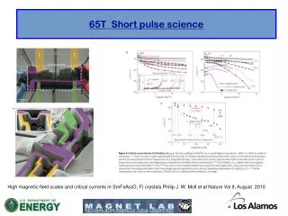
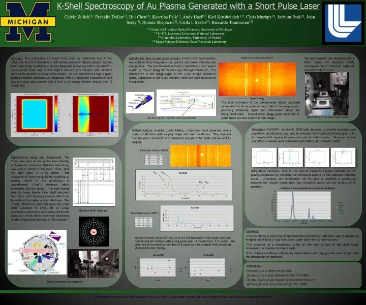
K-Shell Spectroscopy of Au Plasma Generated with a Short Pulse Laser Calvin Zulick[1], Franklin Dollar[1], Hui Chen[2], Katerina Falk[3], Andy Hazi[2], Karl Krushelnick[1], Chris Murhpy[3], Jaebum Park[2], John Seely[4], Ronnie Shepherd[2], Csilla I. Szabo[4], RiccardoTommasini[2] [1] Center for Ultrafast Optical Science, University of Michigan [2] L-472, Lawrence Livermore National Laboratory [3] Clarendon Laboratory, University of Oxford [4] Space Science Division, Naval Research Laboratory Abstract: The production of x-rays from electron transitions into K-shell vacancies (K-α/β emission) is a well known process in atomic physics and has been extensively studied as a plasma diagnostic in low and mid Z materials[1-2]. Such spectra from near neutral high-Z ions are very complex and therefore difficult to describe with analytical models. In this experiment a high Z (gold) plasma emission spectrum was measured with a transparent cylindrically bent quartz crystal spectrometer with a hard x-ray energy window ranging from 17 to 102 keV. • Cylindrically Bent Crystal Spectrometer: A hard x-ray spectrometer was used to time-integrate x-ray spectra and prove intensity and energy data. The spectrometer utilized a cylindrically bent quartz crystal to “focus” Bragg diffracted x-rays through a lead slit. The dependence of the Bragg angle on the x-ray energy introduces spatial separation in the x-ray energies which are then resolved on image plate. Image Plate showing Eu Spectra The spectrometer was designed with a 60cm stand off distance which corresponds to a collection angle of approximately 50 mrad. Higher Energy The axial symmetry of the spectrometer allows duplicate information to be recorded on each side of the image plate, providing additional signal and information about the background noise. Second order Bragg peaks from the K-alpha signal are also evident in this image. Spectrometer in the Titan target chamber CAD drawing and schematic of the spectrometer Simulation: FLYCHK[4], an atomic NLTE code designed to provide ionization and population distributions, was used to simulate the K-alpha and K-beta spectra for Au targets with varying temperatures and ionization states. Temperature and ionization estimates were calculated with HYADES (a 1-D hydro-code). K-Shell Spectra:K-Alpha1,2 and K-Beta1,2 transitions were observed over a series of 40 shots with varying target and laser conditions. The observed spectra were consistent with tabulated energies[3] for both cold Au and Eu targets. Tabulated Values (NIST) Experimental Setup and Background:The Titan laser, part of the Jupiter Laser Facility at Lawrence Livermore National Laboratory, was used to deliver a 350 joule, 10 ps, 1054 nm laser pulse to a Au target. The absorption of laser energy by the resulting Au plasma results in the production of suprathermal (“hot”) electrons which propagate into the target. The high energy electron beam knocks inner shell electrons from their orbit leaving vacancies which can be replaced by higher energy electrons. The energy released as electrons relax into inner shell vacancies is given off as x-rays (commonly referred to as K-alpha and K-beta radiation) which differ in energy depending on the original shell position of the electron. Using these estimates, FLYCHK was used to establish a better estimate on the plasma conditions by matching the simulated spectra to the observed intensity ratios. Ultimately, this information will be used to establish a connection between the plasma temperature and ionization states and the production of positrons. Kα2 Kα1 Kβ1 Kβ2 Electron Shell Diagram Tabulated Values (NIST) • Summary: • The cylindrically bent crystal spectrometer provides an effective way of measuring K-alpha and K-beta x-rays from short pulse laser-matter interactions. • The presence of a nanosecond pulse on the rear surface of the gold target increased the K-alpha to K-beta ratio. • The plasma conditions inferred by the K-shell x-rays may provide some insight into the production of positrons. We performed a series of shots in which the backside of the target was pre-heated and pre-ionized with a long pulse laser (3 nanosecond, 1-10 joule). We observed an increase in the ratio of K-alpha to K-beta signal with increasing short pulse laser energy. References: [1] Kneip, S. et al. HEDP 4 41-48 (2008) [2] Jiang, Z. et al. Phys. Plasmas 2 5 1702-1711 (1995) [3] http://www.nist.gov/physlab/data/xraytrans/index.cfm [4] Chung, H. et al. http://nlte.nist.gov/FLY/ (2008) Titan Experimental Chamber This work performed under the auspices of the U.S. DOE by LLNL under Contract DE-AC52-07NA27344 and was funded by LDRD #10-ERD-044