Download
1 / 24
250 likes | 699 Views
Lectures mycoplasma,ureaplasma spirochetes-borrelia, leptospira,treponema chlamydia, rickettsia Mycobacterium att. Spirochetales Genus : TREPONEMA, BORRELIA, LEPTOSPIRA,

E N D
Lectures mycoplasma,ureaplasma spirochetes-borrelia, leptospira,treponemachlamydia, rickettsiaMycobacterium att
Spirochetales Genus : TREPONEMA, BORRELIA, LEPTOSPIRA, Species: Treponema pallidum – spp.pallidum - syfilis, -ssp.pertenue, ssp.endemicum Treponema carateum – pinta Borrelia recurrentis – epidemic reccurent fever Borrelia species. – endemic recurent fever Borrelia burgdorferi – borreliosis Leptospira interrogans, L. icterohemorhagicus ...... – leptospirosis Common properties – helical gram negative, not cultivable
Treponema Morphologically identical common serological respond in man – (VDRL, FTA), PNC susceptibility Different epidemiology and clinical signs T.pallidum ssp.pallidum – veneric disease syfilis T.pallidum ssp.endemicus – bejel, T.pallidum ssp. pertenue – framboisie, T.carateum – pinta Physiology and structure - Tr.pallidum „ Spirochete “ = curled hair, strictly human pathogen not cultivable Slow replication (time of replication = 30 hrs) Too thin to be visible in light microscope ( darkfield, fluorescence) non veneric
Patogenesis and immunity OMP – outer membrane protein – adherence, hyaluronidase – only virulent – perivascular infiltration. Host cells are covered by fibronectin – produced by T.p. – anti phagocytal properties Immune answer of patient = tissue destruction Clinics – 3 phases 1) skin in the place of penetration – ulcus durum – primary site of replication (multiple T.sp in ulcus ) endarteritis a periarteritis, infiltration of ulcer with PMNL and makrophages, fagocytosis – spirochets are able to survive it 2) disseminated disease – generalised skin lesions multiple T.sp in blood 3) Late manifestation in any organ or tissue Spontanneous remmission possible
Laboratory dg Microscopy – early dg - in darkfield in the 1st., 2nd., stadium and congenital syfilis – not appropriate for oral lesion – not pathogenic spirochetes - fluorescence, - impregnation with silver Cultivation – not cultivable on artificial media Serological diagnosis – non treponemal tests – IgG or IgM antibodies cross reactivity with antibodies against lipid ( antigen ) from destructed cells cardiolipin - VDRL, RRR test – floculation of cardiolopin antigen with patien sera with antitreponemal antibodies, VDRL j the only appropriate test for CSF testing and for control of efficiency of therapy - treponemal tests – specific, confirmation FTA-ABS fluorescence absorption test, TPHA – treponema pallidum hemagglutination test
False positive Not treponemal– viral infections, colagenosis of vascular system, gravidity, recent immunisation, IM,application of feroin, lepra, malaria Treponemal – pyodermia, skin neoplasma, akne, mycosis, ulcus cruris, reumatoid arthritis, psoriasis, SLE, gravidity, herpes genitalis, autoimmune diseases Sensitivity of test in non treated syphilis primary secundary latent late VDRL 59-87% 100% 73-91% 39-94% FTA 86-100 99-100 96-99 96-100 TPHA 64-87 99-100 96-100 94-100% Interpretation of serological testsNontreponemal tests positive later in the 1 st stadium ,present during not treated lues during the 2nd stadium, 25% of not treated are negat. in the 3rd st.., appropriate for therapy control Treponemal tests less influenced by therapy, specificity 98% Positive in newbornes of inficated mothers ? Congenital syphilis or passive transfer of maternal antibodies
Therapy PNC (!Herxheimer reaction! , V PNC for later stadia G PNC for congenital and late, TTC, ERY, CMP – in the case of allergy, for neurosyphilis only PNC or CMP Prevention and control vaccine is not available, therapy of sexual partners, promiscuity a iv drug abusers, AIDS Epidemiology world wiede spread - direct contact - congenital - transfussion of blood Infectivity – low (30%), Infection of fetus soon after infection of mother-bacteraemia in early stages – not treated mother – spontanneous repeated bacteraemias 8 years
Other treponematoses T.pallidum ssp.endemicum – bejel, endemic syphilis, interhuman transmission, nonveneric disease 3 stages: inicial oral lesions, sec. oral defects late gumatas T.pertenue – framboisie (yaws) – granulomatouse disease direct contact with ill skin eatly stage – skin late stage– destruction od skin, lymphatic nodes and bones T. carateum – pinta – primary skin Small papuls, spreading, persisting for years reccurent, disseminating hypopigmentation Therapy: PNC, TTC, CMP Dg: clinical, Mi from lesions, VDRL
Geografical spread of bejel (blue), framboisie (red) a pinta (yellow
BorreliaB.recurrentis- Reccurent fever – epidemic - endemic B.burgdorferi – Lymes diseases– borreliosis – 1977 typical morphology of spirochetales faible staining (anillin stain), flagella, motile, Not cultivable nutritional requirements, replication time 18 hrs, Lab.diagnostic: *Mi in blood smear(reccurent fever), ...........biopsie of skin (borreliosis) *serologic (borreliosis) – IgM, IgG, CSF – Ab index
Borrelia burgdorferi–Lyme disease – borreliosis 1977 arthriis in children tick borne - Ixodes ricinus... (spring, fall differeent species of borrelia - B. garini, B.afzeli... different clinical signs present in skin, isolation in later stages (? symptoms – caused by living borrelia or immunopathological mechanismus?) Clinics: INKT 3-30 days, 1) erytema chronicum migrans +nonspecific signs - 4 weeks 2) late manifestation (80% non treated) *neurological and cardial symptoms *arthralgies a arthritis Dg : Mi – scarecly serology and clinical (ECM ) Therapy:PNC – longtime, DOXY, AMOX, in combination
Serology – only in positive history and clinicIFT ,ELISA tests – all stages Nontreated: IgM 2-4 weaks after appearence of ECM , (MX 6-8w), longlasting in persistent infection IgG MX after 4-6 months., persistent during late manifestation Radical and early threatment: - no IgG formation dg neuroboreliosis – detection of Ab in CSF – antibody index – detection of intrathecal production of IgG ab IgG posit. in CSF can be trasfered pasively from blood via HE barriere, IgM cannot be – pentamer molecules Confirmation – Western blot – detection of antibodies against unique antigens (41 kD – flagellar – IgM., 31kD OspA, 34kD OspB, 60kD – in later stages, IgG DNA amplification PCR
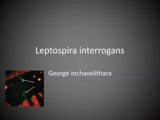
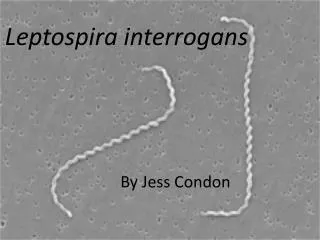
Leptospires L.interrogans ? – 19 serogroups and 172 serotypes (ssp.icterohemorhagica) L.biflexa – 38 serogroups and 65 serotypes Pathogennic for annimals spread by urine, canals – surviving 6weeks 22 serotypes responsible for human diseases (diseases are not serotype specific) Clinics: subclinical disease, fever, systemic disease (Weil disease) Pathogenesis: Spread via intact mucus and small skin injuries, infection of skin and small vessels and all organs (meningitis, hepatal a renal dysfunction, hemorrhagies) Dg. microscopy, agglutiantion
Mycoplasma 69 serotypes and ureaplasma 3 serotypes Common properties, commonly present in the nature, 3 human pathogens, different clinical entities, ethiopathogenesis not exactly clear Mycoplasma pneumoniae, M. hominis, Ureaplsma urealyticum The smallest bacteria without cell wall living free in nature - PNC resistant, Gram negative, formerly classified as viruses Cultivation - media with sterols, colonies of ox eyes generation time 1-6 hrs, Cross reactitivty (main Ags are proteins and glycoproteins) extracelular pathogens , respiratory infections in summer and fall (epidemies in 4-8 years cycles, most commonly school age children Transmission - inhalation and sexually
Clinical picture: M.pneumoniae – infections of respiratory tract, atypical pneumonia M.hominis- pyelonefritis, pelvicitis, post delivery fevers, Ureaplazma – uretritis Diagnosis: microscopy – faintly stainable – no cell wall cultivation: M. pneumoniae – specific media, ox eyes colonies, medium with glucose – change of color indicator when pH chnged because od metabolism of glc to acid – indication of the growth Ureaplasma – detection by metabolicsm of urea and ATB resistance – rapid test serology : detection of IgM against M. pneumoniae Therapy ERY, TTC – M. pneumoniae M. hominis – resitence for ERY
Chlamydiae • Formelly classified as big viruses because of their i.c. parasitism, they have special growth cycle, do not have peptidoglycan in the cell wall • - both DNA and RNA (they are bacteria), - external and internal membrane like G- (LPS), - they are susceptible to some ATB C. trachomatis, C. psittaci, C.pneumoniae Present in 2 morphological formes: - Elementar body- EB – small, infectious, not dividing - Reticular body -RB – bigger noninfectious, dividing, i.c., metabolical activity – energetic parazitism – can synthetise proteins, cannot produce energy
Chlamydiae – growth cycle • Elementar body (EB) is attached on the surface of the target cell by receptors, internalised by phagocytosis. The cell wall of chlamydia does not allow bacteria to unify the phagosome with lysosome – i.c. surviving. EB is reorganised to reticular body, that can divide by binnary fission – 24 hrs, than reorganised again to EB, the cell crashes and EB are transformed to the living the cell (48-72hrs after infection)
Chlamydia trachomatis • 3 serovars – trachomatis (12 serotypes), LGV (3 serotypes) and annimal • C.trachomatis – A,B,Ba,C – trachom – blindness B, D-K – in women – cervicitis, uretritis, proctitis, conjunctivitic + coomplications – in men uretritis, proctitis, conjunctivitis + complications – in children – conjunctivitis, pneumonia, - infection during delivery – farygeal carriage and presence at GIT • LGV- L1, L2, L3 -Lymfogranuloma venereum + complication • Pathogenesis – enter via minimal injury - direct destruction of epithelial cells + inflammatory reaction. In LGV infection is localised in lymphnodes– abscess, rupture, fissures, fistulas
Clinical presentation,diagnosis, therapy • Trachom – chronical keratoconjunctivitis – strictures, chronical irritation of cornea via the lid that is deformed, panus, blindness – transmission via hands, towel, fleas, carriage in respiratory tract and GIT in children. • Inclussion conjunctivitis- adult - folicular - mucopurulent pus, keratitis, infiltation of cornea – in connection swith earlier genital infection, autoinnoculation • Neonatal conjunctivitis and pneumonia – inoculation at birth during delivery – hyperaemia and copious pus, interstitial pneumonia, rhinitis, not fever, staccato cough • Urogenital infection – sexually transmitted non gonococcal uretritis – mucopurulent discharge, hypertrofic ektopium, in men assymptomatic • Reiter syndrome – uretritis, conjunctivitis, polyartritis, mucockutaneous lessions – younger men – sequaelae of uretritis • Therapy: i.c. – ERY, TTC, AZI
LVG - Chronic sexually transmitted disease, more often in men, primary lession – small no aches discrete. Heeling – headaches, fever, myalgia. Secudarye stadium – inflamtion of lyphnodes – aches, fluctuation, bubo, growing, drenaige via fistules Systemic manifestation – fever, anorexia, meningismus, arthralgia. Chronical LVG – genital ulceration, fistuls, strictures, genital elephantiasi • Laboratory diagnosis – cytology – Giemsa stainning – cultivation – on tissues – detection of antigen (OMP or LPS) – ! In cells! – sampling of epitelial cells – detection of Nucleic acid -PCR – detection of viable and non viable cells – is not usefull for monitoring the success of therapy – serology – in endemic area – IgG positive
C. psittací, C. pneumoniaeChlamydophila • Respiratory infection – transmitted from parrots and spread via RES and blood. Lung infection. Immune response : mostly lymphocytes. – oedema,thickness of alveolar wall, cyanosis, anoxia – involevement of CNS GIT, systemic (rash, carditis, hepatomegalia, keratoconjunctivitis) • Conjunctivitis of children in Thaiwan – pharyngeal carriage – TWAR strains – bronchitis, pneumonia, sínusitis...., atypical pneumonia, • Dg – serological • TH – ERY, TTC
Rickettsiacae • Aerobic, i.c. or e.c. parasits, G-, growing only in eucaryotic cells – formerly described as viruses, surviving phagocytosis, spread to cytoplasma where they divide by binary fission • 4 genuses – Rickettsia, – Coxiella (e.c. survive outside the cell) , no vector – Rochalimea – e.c., – Ehrlichia (forms elementary and initial bodies and morula - all forms are i.c.) • Transmitted by insects – (with exeption of Coxiella)
Rickettsiae • Rickettsia rickettsii –Rocky mountains fever – tickborne – transovarial transmission, reservoir – rodents – spotted fever. Dg – Giemsa staining, tissue clutures - Weil Felix test – cross reactivity with Proteus sp. antigens. Th: TTC, CMP • Rickettsia prowazekií – spotted fever – human louse vector – man is reservoire (bad living conditions) . Louse dies for infection – not transovarial transmission – epidemic typhus – macular exantem. Reactivation after years Brill Zinser´s disease. • Rickettsia typií – endemic typhus – vector :Xenophylla cheopsis, reservoires - rodent . Maculopalar rash. Not complicated. • Ricketsia Tsutsugamushi – Pacific
Coxiella, Ehrlichia, Rochalimaea • C. burnetti – Q fever – inhalation of infectious particules from contaminated environment, no insect vector. Proliferation in respiratory tract – dissemination – pneumonia, granulomatous hepatitis. Infection of wild annimals and insect, spred via urine, milk, stool – raw milk, vegetable Acute infection – rapid development, flu disease, respiratory infection, hepatosplenomegalia. Chronic infection – diffuse granulomatosis – chronic endocarditis. Dg – difficult by serology – antigenic variation od phases I and II • Ehrlichia – Paul Ehrlich – leucocytar rickettsiae, parasitism on lymphocytes, neutrophils a monocytes. Not ery. Ehrlichiosis – tick borne – flu signs, leucopenia, trombocytopenia. Rash only in 20%. Good prognosis • R.quintana, R. henselae