Download
1 / 45
580 likes | 2.21k Views
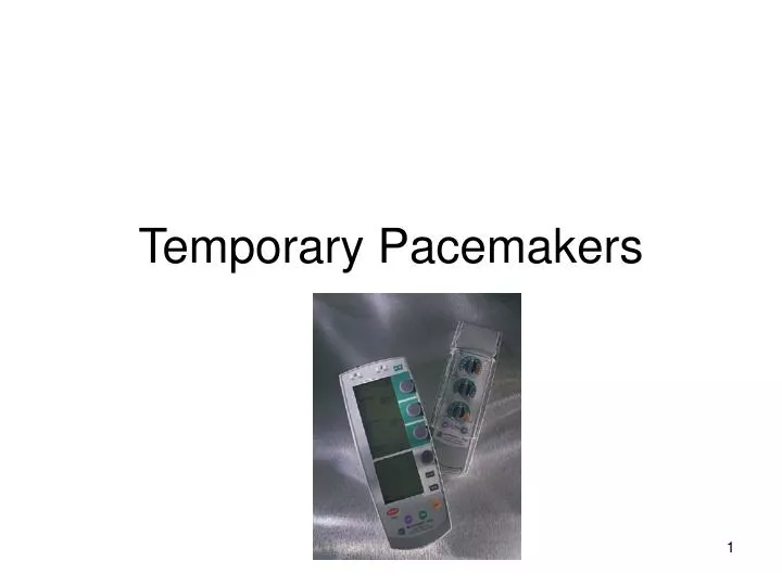
Temporary Pacemakers. Principles of Pacing. Temporary pacing types Transcutaneous Emergency use with external pacing/defib unit Transvenous Emergency use with external pacemaker Epicardial Wires sutured to right atrium & right ventricle Atrial wires exit on the right of the sternum

E N D
Principles of Pacing • Temporary pacing types • Transcutaneous • Emergency use with external pacing/defib unit • Transvenous • Emergency use with external pacemaker • Epicardial • Wires sutured to right atrium & right ventricle • Atrial wires exit on the right of the sternum • Ventricular wires exit on the left of the sternum
Principles of Pacing • Modes of Pacing • Atrial pacing • Intact AV conduction system required • Ventricular pacing • Loss of atrial kick • Discordant ventricular contractions • Sustains cardiac output • Atrial/Ventricular pacing • Natural pacing • Atrial-ventricular synchrony
Principles of Pacing • Commonly used modes: • AAI - atrial demand pacing • VVI - ventricular demand pacing • DDD – atrial/ventricular demand pacing, senses & paces both chambers; trigger or inhibit • AOO - atrial asynchronous pacing
Principles of Pacing • Atrial and ventricular output • Milliamperes (mA) • Typical atrial 5mA • Typical ventricular 8-10 mA • AV Interval • Milliseconds (msec) • Time from atrial sense/pace to ventricular pace • Synonymous with “PR” interval • Atrial and ventricular sensitivity • Millivolts (mV) • Typical atrial: 0.4 mV • Typical ventricular: 2.0mV
Principles of Pacing (cont.) • Atrial/ventricular rate • Set at physiologic rate for individual patient • AV Interval, upper rate, & PVARP automatically adjust with set rate changes • Upper rate • Automatically adjusts to 30 bpm higher than set rate • Prevents pacemaker mediated tachycardia from unusually high atrial rates • Refractory period • PVARP: Post Ventricular Atrial Refractory Period • Time after ventricular sensing/pacing when atrial events are ignored
Principles of Pacing • Electrical Safety • Microshock • Accidental de-wiring • Taping wires • Securing pacemaker • Removal of pacing wires • Potential myocardial trauma • Bleeding • Pericardial effusion/tamponade • Hemothorax • Ventricular arrhythmias
Pacemaker • Medtronic 5388 Dual Chamber (DDD)
Pacemaker ECG Strips • Every pacer spike should have a following p-wave or QRS complex
Normal Pacing • Atrial Pacing • Atrial pacing spikes followed by p waves
Normal Pacing • Ventricular pacing • Ventricular pacing spikes followed by wide, bizarre QRS complexes
Normal Pacing • A-V Pacing • Atrial & Ventricular pacing spikes followed by atrial & ventricular complexes
Normal Pacing • DDD mode of pacing • Ventricle paced at atrial rate
Stimulation Threshold testing • Stimulation threshold (output) • Definition: Minimum current necessary to capture & stimulate the heart • Testing • Set pacer rate 10 ppm faster than patient’s HR • Decrease mA until capture is lost • Increase output until capture is regained (threshold capture) • Output setting to be 2x’s threshold capture • Example: Set output at 10mA if capture was regained at 5mA
Sensitivity Threshold Testing • Set pacer rate 10 ppm slower than patient’s HR • Increase sensitivity to chamber being tested to minimum level (0.4mV) • Decrease sensitivity of the pacer (↑mV) to the chamber being tested until pacer stops sensing patient (orange light stops flashing) • Increase sensitivity of the pacer (↓mV) until the pacer senses the patient (orange light begins flashing). This is the threshold for sensitivity. • Set the sensitivity at ½ the threshold value. Example: Set sensitivity at 1mV if the threshold was 2mV
Failure to CAPTURE • Atrial non-capture • Atrial pacing spikes are not followed by P waves
Failure to CAPTURE • Ventricular non-capture • Ventricular pacing spikes are not followed by QRS complexes
Failure to CAPTURE • Causes • Insufficient energy delivered by pacer • Low pacemaker battery • Dislodged, loose, fibrotic, or fractured electrode • Electrolyte abnormalities • Acidosis • Hypoxemia • Hypokalemia • Danger - poor cardiac output
Failure to CAPTURE • Solutions • View rhythm in different leads • Check connections • Increase pacer output (↑mA) • Change battery, cables, pacer • Reverse polarity
Failure to SENSE • Atrial undersensing • Atrial pacing spikes occur irregardless of P waves • Pacemaker is not “seeing” intrinsic activity
Failure to SENSE • Ventricular undersensing • Ventricular pacing spikes occur regardless of QRS complexes • Pacemaker is not “seeing” intrinsic activity
Failure to SENSE • Causes • Pacemaker not sensitive enough to patient’s intrinsic electrical activity (mV) • Insufficient myocardial voltage • Dislodged lead • Electrolyte abnormalities • Low battery • Malfunction of pacemaker
Failure to SENSE • Danger – potential (low) for paced ventricular beat to land on T wave
Failure to SENSE • Solution • Increase pacemaker’s sensitivity (↓mV) • View rhythm in different leads • Check connections • Reverse polarity • Change cables, battery, pacemaker • Check electrolytes
Oversensing • Causes • Pacemaker inhibited due to sensing of “P” waves & “QRS” complexes that do not exist • Pacemaker too sensitive • Pacemaker failure • Danger - heart block, asystole
Oversensing • Solution • Decrease pacemaker sensitivity (↑mV) • View rhythm in different leads • Check connections • Change cables, battery, pacemaker • Reverse polarity
Competition • Assessment • Pacemaker & patient’s intrinsic rate are similar • Unrelated pacer spikes to P wave, QRS complex • Fusion beats
Competition • Causes • Asynchronous pacing • Failure to sense • Mechanical failure: wires, bridging cables, pacemaker • Loose connections • Danger • Impaired cardiac output • Potential (low) for paced ventricular beat to land on T wave
Competition • Solution • Assess underlying rhythm • Slowly turn pacer rate down • Increase pacemaker rate • Troubleshoot as for failure to sense
Assessing Underlying Rhythm • Carefully assess underlying rhythm • Right way: slowly decrease pacemaker rate
Assessing Underlying Rhythm • Assessing Underlying Rhythm • Wrong way: pause pacer or unplug cables
Answers • AAI: normal atrial pacing • Sinus rhythm: no pacing; possible back-up setting AAI, VVI, DDD • DDD: failure to sense ventricle; increase ventricular mA • VVI: ventricular pacing • DDD: failure to capture atria or ventricle; increase atrial & ventricular mA • DDD: normal atrial & ventricular pacing • DDD: normal atrial sensing, ventricular pacing • DDD: oversensing; decrease ventricular sensitivity