Download
1 / 46
651 likes | 2.76k Views
BRONCHOPNEUMONIA PREPARED BY , M. HASEENA ER DEPT. DR . AHMAD ABANAMY HOSPITAL. NURSING CASE STUDY OF A PATIENT WITH BRONCHOPNEUMONIA. Demographic data NAME :X AGE : 9 YEARS SEX : MALE NATIONALITY :TURKISH DATE OF ADMISSION :12/01/13

E N D
BRONCHOPNEUMONIAPREPARED BY , M. HASEENA ER DEPT. DR . AHMAD ABANAMY HOSPITAL
NURSING CASE STUDY OF A PATIENT WITH BRONCHOPNEUMONIA Demographic data NAME :X AGE : 9 YEARS SEX : MALE NATIONALITY :TURKISH DATE OF ADMISSION :12/01/13 CHIEF COMPLAINTS :fever,COUGH , SOB. DIAGNOSIS : BRONCHOPNEUMONIA
PHYSICAL ASSESSMENT : GENERAL APPEARANCE Child is looking dull respiratory distress present, wheezing present, skin is warm to touch Vital signs: Temperature : 38.8°c Heart rate : 115 /mnt, Respiration : 54 b/ mnt nasal flaring present Blood pressure : 100/ 80 mmHg Spo2 : 88 % in room air GENERAL MEASUREMENT Head circumference – 44cm Chest circumference –28cm Weight -33kg Length -110cm
PHYSICAL ASSESSMENT SKIN • Normal skin colour • Hair soft and silky • Warm to touch • Nails to end of fingers and often extend NOSE • Nostrills patent bilaterally • Nasal flaring present • Nasal discharges present
PHYSICAL ASSESSMENT MOUTH AND THROAT • Uvula midline • Secretion present • Tongue moves freely • Gag reflex present • Teeth is normal in colour • Productive cough present NECK • Short neck present • Turns side to side easily • No lymph node enlargement present
PHYSICAL ASSESSMENT CHEST • Bilateral chest movement present • Nipple is symmetrical • Retraction present • Crackles present • Decreased breath sound present • Tachycardia present ABDOMEN • Soft to palpate • Umbilicus is normal • Bowel sound is normal on auscultation
PHYSICAL ASSESSMENT GENITALIA • Urinary meatus at tip of glans penis • Palpable testes in scrotum and is normal in shape • Adequate voiding and defecation present BACK • Spine is intact • No spinal deformity present EXTREMITIES • Full range of motion present • Ten fingers and ten toes present • Nails are normal in shape and colour
ABBREVIATION OF VACCINES • Hep B : Hepatitis B • RV : Rotavirus • DPT : Diphtheria , Pertuses, Tetanus • HiB : Haemophilus influenza type B • PCV : Pneumococcal vaccine • IPV : Inactivated poliovirus • MMR : Measeles, Mumps, Rubella • Hep A : Hepatitis A • MCV 4 : Meningococcal virus
PATIENT HISTORY Past Medical History: patient Xs is known case of bronchial asthma since childhood. And he is on medication (nebulization) , and no other treatment . Present medical history: patient xs is came to ER Dept due to the complaints of high grade fever, severe cough, since 2 days. Shortness of breath , poor oral intake since one day. Seen and examined by our ER Paediatrition, nebulisation with ventolin, atrovent and pulmicort given. Inj . hydrocortisone 100mg IV given .But no improvement so the patient is admitted to ward for further conservative management Surgical history : patient xs has no present and past surgical history
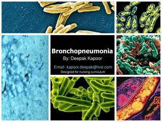
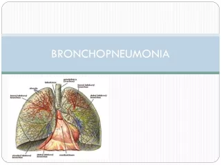
TOPIC PRESENTATION • Bronchopneumonia is a severe type of pneumonia that is characterized by multiple areas of acute and isolated consolidation that affect one or more pulmonary lobes. It is one of the most serious infection in childrens.The disease assumes alarming proportion if both the lungs are affected. Great care has to be taken if the patient suffers from bronchopneumonia. If it is left untreated, the outcome may be fatal.
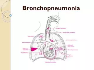
ANATOMY AND PHYSIOLOGY OF RESPIRATORY SYSTEM • The respiratory system is situated in the thorax, and is responsible for gaseous exchange between the circulatory system and the outside world.
ANATOMY AND PHYSIOLOGY The respiratory system is represented by the following structures THE NOSE • It consist of the visible external nose and the internal nasal cavity. The nasal septum divide the nasal cavity into right and left sides. Air enters two opening , the external nares (nostrils and naris ) and pasess into the vestibule and through passages called meatuses. The bony wall of the meatus called concha , are formed by the facial bone ( the inferior nasal concha and the ethmoid bone ) . from the meatuses the air then funnels into left and right internal nares. Hair , mucus, blood capillaries and cilia that lines the nasal cavity filter, moisten ,warm and eliminate debris from the passing air .
ANATOMY AND PHYSIOLOGY PHARYNX : The pharynx ( throat ) consist of the following three region , listed in order through which incoming air passess NASOPHARYNX : It receives the incoming air from the two internal nares , the two auditory tubes that equalize the air pressure in the middle ear also enter here . the pharyngeal tonsils ( adenoid ) lies at the back of the nasopharynx. OROPHARYNX : It receives air from the nasopharynx and food from the oral cavity , the palatine and lingual tonsils are located here . LARYNGOPHARYNX : It passess food to the oesophagus and air to the larynx
ANATOMY AND PHYSIOLOGY THE LARYNX : It receives air from the laryngopharynx . it consist of several piece of cartilage that are joined by membranes and ligaments . EPIGLOTTIS It is the first piece of cartilage of the larynx , is a flexible flap that covers the glottis . the upper region of the larynx , during swallowing to prevent the entrance of the food . THYRIOD CARTILAGE It protect the front of the larynx , a forward projection of this cartilage appears as the ADAMS apple ( laryngeal prominence ) .
ANATOMY AND PHYSIOLOGY The upper vestibular folds ( false vocal cords ) contain muscle fibres that brings the folds together and allow the breath to be held during periods of muscular pressure on the thoracic cavity ( eg : straining while defecating , or lifting a heavy object ) The lower vocal folds ( true vocal cords ) contain elastic ligament that vibrate when skeletal muscle move them into the path of out going air . various sound including speech are produced in this manner . CRICOID CARTILAGE These are supporting the larynx
ANATOMY AND PHYSIOLOGY TRACHEA The trachea ( wind pipe )is a flexible tube about 10-12 cm long and 2.5 cm in diameter The mucosa is the inner layer of the trachea contain mucus producing goblet cells and pseudo stratisfied ciliated epithelium . the movement of the cilia sweeps debris away from the lungs towards the pharynx . The submucosa is a layer of areolar connective tissue that surround the mucosa . The adventitia is the outermost layer of the trachea . it consist of areolar connective tissue . LUNGS The lungs are a pair of cone shaped bodies that occupy the thorax , the mediastenum , the cavity containing the heart , separate the two lungs . left and right divided by the fissure into two and three lobes . each lobe is further divide d into lobules with terminal bronchioles . blood vessels , lymphatic vessels and nerves penetrate each lobe .
ANATOMY AND PHYSIOLOGY The lungs are the sites for gaseous exchange, and are situated within the thoracic cavity. They occupy 5% of the body volume in mammals when relaxed., and their elastic nature allow them to expand and contract with the process of inspiration and expiration. Pleura is a double layered membarane consisting of an inner pulmonary ( visceral ) pleura which surround each lung . the narrow space between the two membarane is the pleural cavity is filled with pleural fluid , a lubricant secreted by the pleura . Each lung has the following superficial features The apex and the base identify the top and bottom of the lung The costal surface of each lung borders the ribs On the medial ( mediastenal surface ) where each lung faces the other lung , the bronchi , blood vessels, and lymphatic vessels enter the lungs at the hilus .
ANATOMY AND PHYSIOLOGY The primary bronchi are two tubes that branch from the trachea to the left and right lungs . Inside the lungs , each primary bronchus divides repeatedly into branches of secondary ( lobar ) bronchi , tertiary ( segmental ) bronchi , and numerous bronchioles , including terminal bronchioles and respiratory bronchioles . the wall of the primary bronchi is constructed like the trachea , but as the branches of the tree get smaller . the cartilaginous rings and the mucosa are replaced by smooth muscle . ALVEOLAR DUCTS These are the final branches of the bronchial tree . each alveolar ducts has enlarged bubble like swelling along its length . each bubble is called alveolus . some adjacent alveoli are connected by alveolar pores . The respiratory membrane consist of the alveolar and capillary walls . gas exchange occurs across these membarane .
ANATOMY AND PHYSIOLOGY The characteristics are TYPE 1 CELLS : are thin , squamous epithelial cells that constitute the alveolar wall . oxygen diffusion occurs across these cells . TYPE 2 CELLS : These are cuboidal epithelial cells that are interspersed among type 1 cells . it will secrete pulmonary surfactant that reduce the surface tension of the moisture that cover the alveolar walls . a reduction in surface tension permit oxygen to diffuse more easly into moisture . a lower surface tension also prevent the moisture on opposite wall of an alveolus , alveolar duct from cohering and causing the airway to collapse . ALVEOLAR MACROPHAGE Alveolar macrophage cells ( dust cells ) wanders among the other cells of the alveolar wall , removing debris and micro organisam . a dense network of capillaries surround each alveolus . the capillary wall consist of endothelial cell surrounded by a thin basement membarane . the basement membarane of the alveolus and the capillary are often so close that they fuse .
MECHANISM OF BREATHING Breathing occurs when the contraction or relaxation of muscle around the lungs changes the total volume of air within the air passages , ( bronchi , bronchioles ) inside the lungs . when the volume of the lungs changes , the pressure of the air in the lungs also changes . if the pressure is greater in the lungs than out side the lungs , the air rushes out . if the opposite occurs , the air rushes in . INSPIRATION PHASE Inspiration occurs when the inspiratory muscle that is the diaphragm and the external intercostals muscle contract , the contraction of the diaphragm causes an increase in the size of the thoracic cavity , while contraction of the external inter costal muscle elevate the ribs and sternum . thus both muscle causes the lungs to expand , increasing the volume of their internal air passages . in response the air pressure inside the lungs decreases below that of air outside the body . because gases moves from region of high pressure to low pressure , air rush into the lungs .
MECHANISM OF BREATHING EXPIRATION PHASE It occurs when the diaphragm and external intercostals muscle relax . in response , the elastic fibres in lung tissue cause the lung to recoil to their original volume . the pressure of the air inside the lungs then increases above the air pressure out the body and air rushes out .
ETIOLOGY Bronchopneumonia is caused by viruses, bacteria , fungi protozoa and myco plasma Bacteria • Streptococcus • Staphylococcus • Hemophilus influenza • Klebsella Virus • legionella pneumonia Fungi • candidaalbicans Other predisposing factors include: • common in hospitalized patients • its occur as a complication of some other diseases , eg: in children – diphtheria, measles, and whooping cough • In adults- influenza, typhoid and paratyphoid fever • its caused by organism aspirated from mouth
SIGNS AND SYMPTOMS High grade fever • Any body temperature that goes above 37 °c is considered as fever . in bronchopneumoniamfever may be he symptoms for having the disease, especially if it is accompanied by other symptoms such as cold , cough and difficulty of breathing Frequent and excessive coughing accompanied by mucus • Cough is a natural reaction of the body to the presence of certain elements that may irritate the throat. However if coughing may become pesistant and accompanied by mucus , then it’s a sign of something more serious than normal coughing. A person with bronchopneumonia experience frequent and excessive coughing sometime accompanied by mucus. Chest pain • The persons experience difficulty of breathing and also sensation of not getting enough air , as a result the person gasping for air frequently • Fatigue • Irritability • Decreased apetite • Decreased breath sound on auscultation • Headache
PATHOPHYSIOLOGY When bacteria infect the pulmonary lobes, the lungs produce mucus that fills the alveolar sacs. this will cause a condition known as consolidation, which occurs when the lungs fill with mucus, lead to reduce in air space. This reduction in air space makes breathing difficulty causing shortness of breath and labored or shallow breathing
PATHOPHYSIOLOGY VIRUS ENTER THE RESPIRATORY TRACT INFLAMMATION ACCUMULATION OF BRONCHIAL SECRETION ALVEOLI COLLAPSE NARROWING OF AIRWAYS SOB & DOB BRONCHOPNEUMONIA
INTERVENTIONS • Perform comprehensive assessment • Auscultate breath sound , noting areas of decreased or absent ventilation • remove secretions by encouraging coughing • Regulate fluid intake to optimize fluid balance and liquefy secretions • Administer oxygen if hypoxemic • Administer medication as prescribed
DIAGNOSIS • Auscultation of breathing pattern • Chest xray • CBC, Sputum culture , c- reactive protein
TREATMENT • Advise to drink plenty of fluid • Enough rest • Elevate the head of the bed to minimize respiratory effort • Administer oxygen, if needed • Use antibiotics as prescribe • Antipyretics as ordered
COMPLICATIONS • Pleural damage leads to pleural effusion, pleural empyema • Cardiovascular disease • Respiratory deficiency • Acute renal insufficiency in dehydration • Septic distribution of the pneumonia agents through the blood with the development of otitis, meningitis, brain abscess, endocarditis
PRIORITIZATION OF NURSING PROBLEMS • Ineffective airway clearance related to accumulation of trachea bronchial secretion • Hyperthermia related to the inflammatory process • Impaired gas exchange related to inflammation of airways and accumulation of sputum • Acute pain related to ineffective comfort measures and inflammation
NURSING HEALTH TEACHING • Follow up the regimen as per order • Frequent hand washing with soap and water or use hand sanitizer • Advise to have healthy diet and adequate rest,that will keep the immune system strong • Advice to cover the mouth while coughing • Follow up to the hospital after finishing the antibiotic course