Download
1 / 16
170 likes | 578 Views
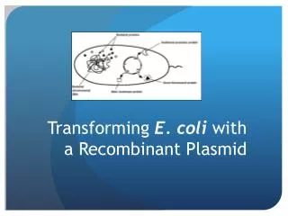
Expression and analysis of recombinant proteins in E. coli. Class 11 CPSC265. Normal cell. Gene (DNA). transcription. mRNA transcript. translation. Protein (amino acids). Structural proteins. Proteins with enzymatic activity. signaling. Cell with recombinant plasmid.

E N D
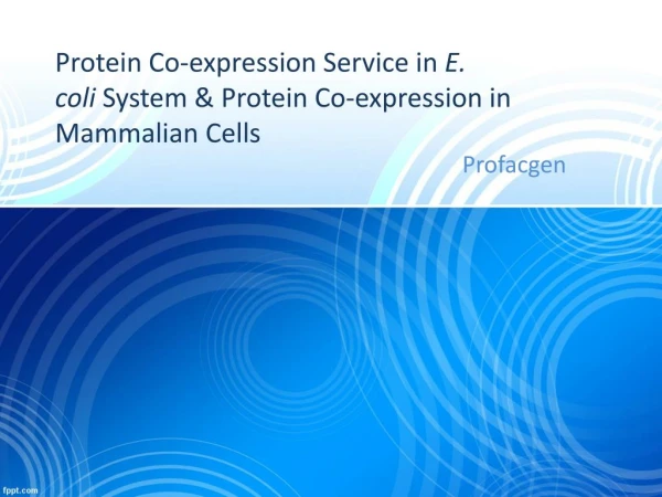
Expression and analysis of recombinant proteins in E. coli Class 11 CPSC265
Normal cell Gene (DNA) transcription mRNA transcript translation Protein (amino acids) Structural proteins Proteins with enzymatic activity signaling
Cell with recombinant plasmid Gene (DNA) transcription mRNA transcript translation Soybean lectin Protein (amino acids) Structural proteins Proteins with enzymatic activity signaling
Inducible gene expression • So that we don’t stop the cells growing (or kill them) we like to grow the cells without our protein being expressed, then switch it on when there are plenty of cells • We do this by manipulating the transcription of the mRNA for the protein from our plasmid
Inducible gene expression (cont) • E. coli naturally keeps some genes turned off. For example, it turns on the genes needed to metabolize galactose, or arabinose, only when these sugars are present. • By cloning the promoter for the operon containing the arabinose or galactose genes in front of our gene in the plasmid, we can keep it turned off until we are ready. Then we add the sugar, and the gene is turned on.
Soy lectin pBAD uses the arabinose promoter
Regulation of pBAD pBAD is regulated by the product of the araC gene (N), a transcriptional regulator that forms a complex with L-arabinose. In the absence of arabinose, the AraC dimer contacts the O2 and I1 half sites of the araBAD operon, forming a 210 bp DNA loop (Figure 1).
What I did already this week • Grew a liquid culture E. coli containing the pBAD plasmid with soybean lectin gene. • When the E. coli cells were abundant, but still growing rapidly, (OD 0.5) I added arabinose to 0.2% concentration • I incubated the “induced” cells for 4 hours
Denaturing SDS protein gels anions of SDS bind to the peptide chain at a ratio of one SDS anion for every two amino acids - Na+ Na+ Na+ Na+ Na+ - - + - - - - - + - - - - - - - - - - Na+ - - - - - Na+ Na+ Na+
In an SDS gel, migration is proportionalto molecular weight One negative forevery 2 a.a.s massivelyoutweighs the nativecharge of the protein Otherwise proteinswould not behave like this. They would migrate towardseither electrode, dependenton pH, compositionand structure
Using acrylamide, we can make a polymer full of hydrophilicpores. The size of the pores is proportional to the % of acrylamide
Stacking gel concentrates proteins into narrow, well-definedbands. Resolving or separating gel resolves them by molecular size.
Coomassie staining • You can view the proteins in the gel by staining with coomassie blue • This is a dye that binds all proteins regardless of their amino acid makeup • Fortunately it is bright blue – no UV required
What you will do today • Take culture and spin down • Extract, denature and reduce disulfide bridges of all proteins • These are done simultaneously by boiling in SDS + beta mercaptoethanol
Today (cont.) • Separate the proteins by size using denaturing, discontinuous SDS electrophoresis • Look for the induced protein using coomassie blue staining