Download

1 / 77
770 likes | 775 Views
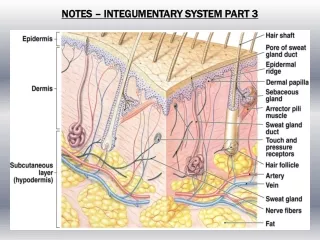
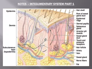
Integumentary System Part 1. The Integumentary system (skin) is made up of several layers: The hypodermis The epidermis The dermis. The H y podermis. This layer is under the dermis and is not considered part of the skin. It serves to separate the skin from the underlying muscle.

E N D
The Integumentary system (skin) is made up of several layers: • The hypodermis • The epidermis • The dermis
The Hypodermis This layer is under the dermis and is not considered part of the skin. It serves to separate the skin from the underlying muscle.
The Hypodermis This layer contains fat tissue and is loosely anchored to the skin. This allows the skin to slide while the fat serves as an insulator and a cushion
The Hypodermis • This layer is the major site of fat accumulation • Distribution differs among the sexes. Its in the thighs and breasts of females and the belly of males
Epidermis • This is the outer most layer and consists of a stratified squamous epithelium. • Four cell types populate it:
Epidermis • This is the outer most layer and consists of a stratified squamous epithelium. • Four cell types populate it: • Keratinocytes • Melanocytes • Epidermal dendritic cells (Langerhan’s cells) • Tactile cells (Merkel cells)
Keratinocytes • These cells produce keratin, a protein made of intermediate fibers.
Keratinocytes • These cells produce keratin, a protein made of intermediate fibers. • These cells attach to each other by desmosomes.
Keratinocytes • These cells are found in the lowest level of the epidermis, the stratum basale (basal cell layer). • These cells divide and give rise to the major epithelial cell type in the epidermis.
Keratinocytes • These cells divide and are pushed towards the upper layers of the epidermis.
Melanocytes • These cells produce melanin and are found occupying the stratum basale.
Melanocytes • These cells produce melanin and are found occupying the stratum basale. • Melanin is stored in intracellular vesicles called melanosomes.
Melanocytes • The melanocytes are “spider” shaped. • The melanosomes migrate to the distal portions of the melanocyte where it is secreted. • The melanin is then taken up by the keratinocytes.
Epidermal dendritic cells (Langerhan’s cells) • These cells originate in the bone marrow and migrate to the epidermis. • They play an important role in the skin’s immune response.
Merkel (tactile) cells • These cells are found at the dermal/epidermal junction (stratum basale) and serve as a touch receptor
Five layers which make up the epidermis. • These are: • Stratum basale • Stratum spinosum • Stratum granulosum • Stratum lucidum • Stratum corneum
Stratum basale (basal layer) • This is the deepest epidermal layer.
Stratum basale (basal layer) • This is the deepest epidermal layer. • Cells in the lowest level undergo constant mitosis in an effort to replace cells lost in the upper layers.
Stratum basale (basal layer) • This is the deepest epidermal layer. • Cells in the lowest level undergo constant mitosis in an effort to replace cells lost in the upper layers. • 10 to 25% of the cells in this layer are melanocytes.
Stratum spinosum (prickly layer) • This layer is several cells thick.
Stratum spinosum (prickly layer) • This layer is several cells thick. • The cells are filled with intermediate fibers which attach to the desmosomes.
Stratum spinosum (prickly layer) • This layer is several cells thick. • The cells are filled with intermediate fibers which attach to the desmosomes. • The desmosomes appear on the cells surface like spikes giving rise to the name prickly.
Stratum granulosum • This layer is 3 to 5 layers thick and is where keratinization occurs
Stratum granulosum • This layer is 3 to 5 layers thick and is where keratinization occurs • Here the cells begin to flatten and the organelles begin to break down.
Stratum granulosum Two types of granules (non membrane bound inclusions) are found in this cell type: • Keratohyaline granules which contain keratin • Lamellated granules contain glycolipids and serve as a water proofing
Stratum lucidum • Seen only where the epidermis is very thick, typically the feet and palms of the hand.
Stratum lucidum • Seen only where the epidermis is very thick, typically the feet and palms of the hand. • It is a histological term for a clear area of cells seen between the stratum granulosum and the stratum corneum.
Stratum corneum (horny layer) • This is the outermost layer and is between 20 to 30 cells thick.
Stratum corneum (horny layer) • This is the outermost layer and is between 20 to 30 cells thick. • It protects against abrasions and is made up of dead cells.
Stratum corneum (horny layer) • This is the outermost layer and is between 20 to 30 cells thick. • It protects against abrasions and is made up of dead cells. • The glycolipids from the lamellated granules waterproof this layer
The Dermis • This layer consists of connective tissue and resident cell types of macrophages, fibroblasts and masts cells.
The Dermis • This layer consists of connective tissue and resident cell types of macrophages, fibroblasts and masts cells. Interspersed in this layer are blood vessels, nerve tissue, hair follicles, oil and sweat glands.
The Dermis The dermis consists of two layers: • Papillary layer • Reticular layer
The Papillary Layer • This consists of areolar connective tissue and lies just below the stratum basale. • The loose nature of this layer allows the cells of the immune system to act quickly on any breach of the epidermal cells
The Papillary Layer • This layer has dermal papillae which project into the epidermis.
The Papillary Layer • This consists of areolar connective tissue and lies just below the stratum basale.
The Papillary Layer • This consists of areolar connective tissue and lies just below the stratum basale. • These carry nerve and capillary networks and touch receptors known as Meissner’s corpuscles to the epidermis through dermal papillae.
The dermal papillae make up the dermal ridges and by extension help form the epidermal ridges. • These are prominent on the hands and feet and form friction ridges and improve gripping. • On the fingers these are known as finger prints.