Download
1 / 28
330 likes | 452 Views
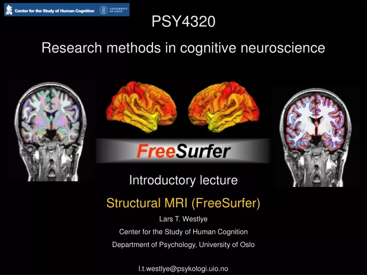
PSY4320 Research methods in cognitive neuroscience. Introductory lecture Structural MRI (FreeSurfer) Lars T. Westlye Center for the Study of Human Cognition Department of Psychology, University of Oslo l.t.westlye@psykologi.uio.no. Cortex (GM). WM. Right. Left.

E N D
PSY4320 Research methods in cognitive neuroscience Introductory lecture Structural MRI (FreeSurfer) Lars T. Westlye Center for the Study of Human Cognition Department of Psychology, University of Oslo l.t.westlye@psykologi.uio.no
Cortex (GM) WM
Right Left T1-weighted MRI volume
Right Left Skull stripped brainmask
White surface Pial surface Pial and white surfaces
Superior temporal gyrus (GM/WM) Right amygdala Right amygdala Left hippocampus Right hippocampus Segmented volume
Pial 3D renderings of the surfaces White
Frontal Parietal Occipital Pial 3D renderings of the surfaces Temporal White
Inflated surface renderings enable visualization of sulci White surface Inflated white surface
Cortical thickness estimation Thickness = distance between pial and white surface at every point
Cortical thickness estimation Approximately 160 000 points (vertices) per hemisphere
Mapping thickness to the surface Warm colors denote thicker cortex
Automatic anatomical parcellation of the surface Precentral Precentral Superior/inferior parietal Superior temporal Middle temporal Superior frontal Middle frontal Entorhinal Fusiform Parahippocampal
Group study: Comparing thickness across subjects Challenge: individual anatomical variability
Individual surfaces Spherical morphing Averaging spheres Mapping back to surfaces Fischl, B.
Averaging across subjects Old Young Young Old Average thickness across 30 young subjects Average thickness across 30 old subjects
Averaging across subjects Average thickness across 30 young subjects Average thickness across 30 old subjects Superior frontal gyrus 2.93 (± 0.15) 2.54 (± 0.17) Young Old
3D renderings of the left hippocampus 2.93 (± 0.15) 2.54 (± 0.17) Automatic segmentation of the hippocampus
3D renderings of the left hippocampus 2.93 (± 0.15) 2.54 (± 0.17) Automatic segmentation of the hippocampus
A few applications Cortical thinning in Alzheimer’s Disease: Thinning typically observed in the medial-temporal and posterior cingulate ”memory network” 2.93 (± 0.15) 2.54 (± 0.17)
A few applications Cortical thinning through the life-span (8-85) Westlye et al. (submitted) 2.54 (± 0.17)
A few applications Cortical thinning through the life-span (8-85) Massive thinning early in life 2.54 (± 0.17) Westlye et al. (submitted)
A few applications Accelerated cortical thinning in healthy adults with increased genetic risk for Alzheimer’s (APOE-4): 2.93 (± 0.15) 2.54 (± 0.17)
A few applications Accelerated cortical thinning in healthy adults with increased genetic risk for Alzheimer’s (APOE-4): 2.93 (± 0.15) 2.54 (± 0.17)
Thicker cortex -> higher P3a peak amplitude Thicker cortex -> shorter P3b latency
Preliminary data shows that regional cortical thickness is negatively correlated with P3a amplitude in children and adolescence (8-18 years of age) Blue areas: Thinner cortex -> higher P3a peak amplitude
A few applications 2.93 (± 0.15) 2.54 (± 0.17) Draganski et al. (2004), Nature
A few applications Cortical thickness changes after intensive Tetris playing 2.93 (± 0.15) 2.54 (± 0.17) Haier et al. (2009), BMC Research Notes