Download
1 / 35
350 likes | 374 Views
Lab 13 Biotechnology: Bacterial Transformation (AP Lab 8). Biotechnology – the use of living organisms or biological systems in research or industrial uses, especially in regard to microorganisms and genetics. Lab 13 Biotechnology: Bacterial Transformation (AP Lab 8)

E N D
Lab 13 Biotechnology: Bacterial Transformation (AP Lab 8) Biotechnology – the use of living organisms or biological systems in research or industrial uses, especially in regard to microorganisms and genetics
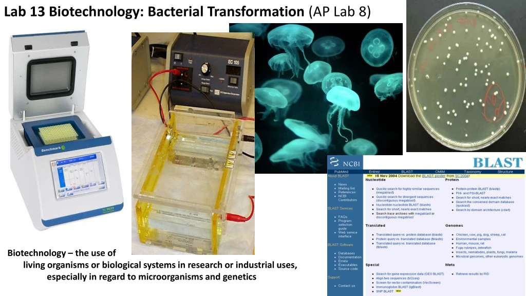
Lab 13 Biotechnology: Bacterial Transformation (AP Lab 8) Title Lab Date Entry in left hand margin (3/12/18) Fill in Table of Contents with info when available Objective(Copy) In this lab students will learn about important technologies and techniques used in biotechnology, including bacterial transformation, gel electrophoresis and polymerase chain reaction. Students will also be introduced to internet research resources used widely in genetics research (BLAST - Basic Local Alignment Search Tool).
Background notes (label section) Our goal is to transform bacteria (E.coli) with a plasmid that includes a gene for making GFP (Green Fluorescent Protein) - the bacterial colonies should than glow under UV light. What is GFP? A protein made in jellyfish that allow them to glow in the dark
Transgenic organisms have genetic material from 2 different species. Why would scientists want to make transgenic organisms of this type? https://www.youtube.com/watch?v=wxf4a4SX84A
GFP is used as an easily detected marker, indicating success of gene insertion process In multicellular organisms the GFP gene can be associated with other genes, showing when and in what tissue the associated genes are turned on
We are going to insert a plasmid with the GFP gene into bacteria cells. Plasmid map
Three things to understand • Transformation process 2. How a plasmid is engineered 3. Arrangement of our genes in the promoter Genes: GFP gene Antibiotic resistance gene
Bacteria will naturally take up plasmid, but we can improve efficiency with a few tricks 1. Cold shock - causes holes to form in membrane 2. CaCl2 in the plasmid solution binds to DNA, negating negative charge and allowing easier passage through holes in membrane (still hydrophobic environment) 3. Cold to hot to cold - rapid temp change induces production of stress proteins that close holes; heat also increases kinetic motion of solution and makes it more likely plasmids will enter holes 4. Back to warmth with nutrient broth to "heal" the bacteria 5. The bacteria are then plated on nutrient agar and allowed to reproduce for 24 hours • What is a typical frequency of transformation in vitro? Transformation efficiency is measured in transformants or colony forming units (cfu) per μgDNA used (# of colonies/ug DNA) Very successful transformation = 1 in 2000 molecules of the plasmid used in transformation
How a plasmid is engineered (how is gene inserted into plasmid?) A plasmid has multiple restriction sites, or places where the plasmid will be cut with restriction enzymes
Restriction enzymes cut DNA at specific sites A plasmid can be cut at a specific site A gene sequence, specifically engineered to have ends compatible with the cut site, can be inserted into the plasmid
Our GFP gene pBAD-GFPuv pBAD = promoter for BAD operon GFPuv = gene for GFP Z, Y and A are genes for making proteins involved in sugar (arabinose) digestion In our plasmid Z,Y and A have been replaced with the GFP gene 717 nucleotides = 238 amino acids Used as a reporter of expression We are using the promoter for an attachment point for polymerase
So we have put the plasmid with the cells, induced transformation, and now we need to incubate the bacteria on nutritive agar so they will grow into identifiably glowing, and therefore transformed, colonies. Under the right conditions these cells will divide every 20 minutes. How many cells will we have in 24 hours, if we only started with one? 3 x 24 = 72, 20-minute increments 272 = 4,722,366,482,869,645,213,696 So what should we find the next day when we look at the plates? Will we be able to identify individual, transformed colonies? (Remember – successful transformation still means low frequency of transformation)
Untransformed bacteria will overgrow our transformants Bacterial lawn Discrete colonies Insert a gene for ampicillin resistance into our plasmid (makes a protein that digests the antibiotic) Then the bacteria are grown on a plate that contains ampicillin - only the transformed bacteria will survive, and since transformation efficiency is low, only a small number of bacteria will survive to make colonies
The U of A Biotech Project (outreach to high schools) provides us with the plasmid, bacteria and other equipment So everything should go perfectly!! But we have to keep in mind that we are working in the dynamic and exciting world of biology, and when working with living systems (as opposed to dull chemistry and physics) things rarely go as planned. I can virtually guarantee that something will go wrong...
3/14/18 In your lab journal date entry and record results in a table (E.coli; 24 hours incubation at 37 dC) Observe our plates… Were we successful??
Oh no!! What happened?? What is the problem? What do we know? What don't we know? What could be possible causes of the problem?
Subtitle “Notes – troubleshooting the problem” What is the problem? We do not have functioning GFP What do we know? Have gene and product for AmpR; we have successful transformation No functional GFP What don't we know? Why don't we have a functional GFP? Is the problem at the gene, mRNA or protein level? Does our plasmid include the gene for GFP? Does the gene make an mRNA? Is the mRNA silenced (kept from translating a protein)? Is the protein functional?
Where could the problem be? GFP Gene -> is gene even there? Plasmid incorrectly engineered? Gene excised by cellular processes? Plasmid not inherited by offspring? -> if gene is present, is it transcribed? Operon regulation? -> what might prevent it from being transcribed? GFP mRNA -> degraded? Silenced in some other way? GFProtein-> degraded? nonfunctional?
Does our plasmid include the gene for GFP? Investigation involves two steps 1. See if there is a gene in our plasmid of the right size, which will “respond” to our primers GFP gene 717 nucleotides = 238 amino acids nucleotide 5’ end sequence --------------------------------------------------------------------------------------------- nucleotide 3’ end sequences nucleotide 3’ end sequence --------------------------------------------------------------------------------------------- nucleotide 5’ end sequences We can investigate this using Polymerase chain reaction and Gel electrophoresis 2. If we confirm #1 above – does this gene have the proper nucleotide sequence? (Was it properly engineered, or did the UofABP mistakenly implant a “mutation” in our gene?) We can investigate this by having the gene we have identified in #1 above sequenced (determine the nucleotide sequence of our gene).
Does our plasmid include the gene for GFP? Step 1 See if there is a gene in our plasmid of the right size, which will “respond” to our primers GFP gene 717 nucleotides = 238 amino acids nucleotide 5’ end sequence ----------------------------------------------------------------------------- nucleotide 3’ end sequences nucleotide 3’ end sequence -----------------------------------------------------------------------------nucleotide 5’ end sequences We can investigate this using Polymerase chain reaction and Gel electrophoresis Subtitle section “Notes on PCR” How can we see if our gene is there? We attempt to “amplify” the gene (PCR) and then see if we can find the copies (GE) Amplify = copy it a bunch of times DNA is amplified by using a process that mimics DNA replication
Polymerase Chain Reaction Polymerase chain reaction multiplies (amplifies) a particular sequence of DNA so that there is enough to detect PCR is a way to artificially replicate a section of DNA, and does so by using some of the biochemical machinery of DNA replication Steps of PCR Cellular or DNA sample is collected and combined with several compounds DNA polymerase - this is a particular type of polymerase with special properties It is able to maintain shape and function at high temperatures Primers that match the 3’ end of the coding and noncoding strand A large concentration of each of the 4 DNA nucleotides (dNTPs) The components are put in a PCR machine https://www.youtube.com/watch?v=JmveVAYKylk
What does PCR do? 1. High temps cause DNA double helix to denature (breaks H bonds) 2. Temps cool allowing primers to bind to 3' end (5' to 3') of exposed coding and noncoding strand of gene 3. Polymerase (Taq) replicates complimentary stands 4. Cycle repeats numerous times to exponentially increase number of gene copies Once we have subjected the genome to PCR we have to see if we amplified anything – was the gene there to amplify? The PCR product is analyzed with gel electrophoresis to determine whether it exists and whether it is the right size.
Uses of PCR Used to amplify the entire genome – DNA fingerprinting Amplify a particular gene of interest Detect the presence of a gene Cross species comparison Genome engineering Detection of viral genes Mass produce a gene Industrial applications
In this lab we wanted to transform bacterial cells… We learned about transformation and the transformation process We successfully transformed cells but no glowing We learned about the PCR process and we used it to see if we could amplify our gene We used gel electrophoresis to see if we had amplified our gene Copy diagram of our gel Subtitle section “Notes – Gel Electrophoresis”
Gel electrophoresis is used to separate DNA fragments Gel electrophoresis is used to separate the DNA fragments into groups according to their size An agar gel is used in gel electrophoresis Agar gel is a porous substance that on a submicroscopic level is honeycombed
The agar is placed on a plate, which is then placed in an electrophoresis chamber
An EC produces a negative charge at the end closer to the depressions and a positive charge at the other end DNA is loaded into the gel DNA is negatively charged, so when the power is turned on, a current will run through the gel and the DNA will "run" towards the positive electrode and away from the negative electrode As the DNA moves down the "lanes" of the gel, different sized fragments will move at different speeds through the tunnels in the gel The electric current is shut off when the smallest fragments reach the end of the gel; fragments are bunched up at different places in the lane according to fragment size The gel is then stained with a dye that binds to DNA
How do you know the size of your fragments? Gel with molecular weight ladder