Download
1 / 43
450 likes | 783 Views
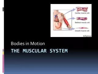
Chapter 7: The Muscular System. Types of Muscle. Skeletal Striated actin/myosin org. voluntary Smooth nonstriated involuntary Cardiac (heart) s triated Involuntary (pacemaker cells). Skeletal muscle. Functions: Movement Posture/Position Soft tissue support Guard entry/exits

E N D
Types of Muscle • Skeletal • Striated • actin/myosin org. • voluntary • Smooth • nonstriated • involuntary • Cardiac (heart) • striated • Involuntary (pacemaker cells)
Skeletal muscle • Functions: • Movement • Posture/Position • Soft tissue support • Guard entry/exits • Maintain body temp. • Voluntary, controlled • Exception of diaphragm • Composition: • Skeletal muscle tissue • Connective tissue • Nerves & Blood vessels • Integrated within the connective tissue layers to deliver nutrients/stimulate cells (axons from CNS) • Very active muscle cells need oxygen/nutrients and waste disposal
Gross anatomy • 3 layers of connective tissue: • Epimysium • Epi = on, mys = muscle • Surrounds entire muscle • Perimysium • Peri = around • Surround fascicles • Endomysium • Endo = inside • Inside fascicle • Muscle fibers = multi-nucleated elongated cells • Fascicle: bundle of muscle fibers
Microanatomy: Inside the muscle cell • Sarcolemma • “sarkos” = flesh • “lemma” = husk • Cell membrane • Sarcoplasm • Cytoplasm • Transverse tubules • “T tubules” • Function in muscle contraction • Myofibril: bundles of myofilaments
Myofibril • Contains myofilaments • Thick and Thin Protein filaments • Actin & Myosin proteins • Actin (Thin) • Myosin (Thick) • Attached to sarcolemma at each end of cell (contraction) • Figure 7-2 (p. 188)
ENERGY Mitochondria and glycogen granules scattered throughout the cell. Cellular respiration: sugar energy ATP for contractions
Microanatomy • Sarcoplasmic Reticulum (SR) • Form of smooth ER • Tubular network around each myofibril • Terminal cisternae “chambers” on SR, sandwich T tubules • Contain high [C] of calcium ions, which are released into sarcoplasm during contraction.
sarcomere • Smallest functional unit of the muscle fiber • Organized, repeating units of myofilaments (thick/thin) • ~10,000 sarcomeres end to end in each myofibril • Banded (striated) appearance from myofilament organization, sarcomeres side by side • Z lines • Boundaries of sarcomere, one sarcomere from Z to Z • M line • Protein that connects neighboring thick filaments • A band (dArk) • Region of thick filaments (some thin in overlap, but more thick) • I band (lIght) • Region of thin filaments
Thick and thin filaments • Thin filaments: twisted strand of actin • Actin • Active sites (reacts with myosin) • Tropomyosin (cover active sites @ rest) • Troponin (stability) • Thick filaments • Myosin • Head and tail • Heads attach to actin during contraction • Calcium is the KEY!
Sliding filaments and cross-bridges • Sliding filament theory • Myosin head (thick) binds to active sites on actin (thin) • Needs calcium to bind to troponin, which moves tropomyosin and exposes active sites on actin
Sliding filaments and cross-bridges • Sliding filament theory • When a muscle contracts: • I band gets smaller • Z lines move closer • Zones of overlap increase • Width of A band stays the same • Thin filaments slide toward center • Thick filaments stay in place • Cross-bridges: myosin heads connected to active sites on actin • “attach, pivot, detach, and return” • (Pulling a rope with one hand)
Neuromuscular junction • Intercellular connection b/t nerves and muscle cells • Muscle fiber (cell)—motor neuron • Axons attach to perimysium and forms synaptic terminal • Synaptic vesicles (ACh) • Synaptic cleft • Motor end plate (binding sites for ACh) • Acetylcholinesterase (AChE) • Breaks down ACh
1. Neuron 2. Sarcolemma (or motor end plate) 3. Synaptic Vesicle 4. Synaptic cleft 5. Mitochondria
SLIDING FILAMENT THEORY (Fig. 7-5) • Active site exposed • Myosin Cross-bridge • Myosin pivots toward center (ADP + P) • New ATP binds, mysin detaches • Myosin “re-primed” when ATP ADP + P
Muscle mechanics • Tension: active force created when muscle fibers contract “pull” • Resistance: passive force that opposes movement • Compression: opposite of tension, “push” • Muscles can only contract (shorten/create tension) • We are not Mr. Fantastic!
Muscle fiber stimulation • Stimulus-Contraction-Relaxation Sequence • Twitch: single sequence • Latent Period: ~2msec, beings at stimulus, action potential propagates across sarcolemma, Calcium released • Contraction Phase: Cross bridges attach, maximum tension ~15msec • Relaxation Phase: Cross bridges detach, calcium reabsorbed by SR, ~25msec
Muscle fiber stimulation • Summation • Addition of multiple twitches, from multiple stimuli • Incomplete Tetanus • Maximum tension not reached (ex. Normal muscle movements) • Complete Tetanus • Maximum tension, fast action potentials, no relaxation phase
Motor unit • Motor unit: All muscle fibers controlled by a single motor neuron • Depends on level on control needed • Eye: 2-3/nerve • Leg: thousands/nerve
Muscle tone & Atrophy Atrophy: smaller and weaker muscles, not stimulated by nerves often Muscle Tone: resting tension
Types of contraction • Isotonic: tension rises, muscle length changes • “Equal tension” • Tension stabilized until relaxation • Isometric: length does not change, tension < resistance • “Equal measure” • (Ex. Pushing a door, picking up a car, sitting, standing)
Muscle elongation • Remember muscles ONLY contract • Return to resting state • Elastic Forces (connective tissues) • Contraction of opposing muscles (Biceps/Triceps) • Gravity (Ex. Stand on one leg)
Muscle energetics • Muscle cells need energy from ATP. • ATP is produced by cellular respiration, an aerobic process (needs oxygen) • Lactic acid production during intense muscle activity, no time for oxygen to diffuse into muscle cell • Muscle fatigue: caused by exhaustion of energy reserves or lactic acid buildup • Lactic acid lowers pH, muscles cannot function normally • Recovery period: muscle returns to preexertion levels • Oxygen debt-breathing rate and depth increase • Lactic acid glucose • Heat Loss: regulates body temperature (ex. Shivering)
Muscle performance • Force: maximum tension • Endurance: duration of activity • Two types of skeletal muscle fibers: • Fast (fast-twitch) • 0.01 sec stimulus response • Large diameter, amount of myofibrils and glycogen reserves • Prone to fatigue • Slow (slow-twitch) • Half diameter of fast, 3x longer stimulus reaction • Longer endurance • Oxygen supply from capillaries • Oxygen storage in myoglobin (oxygen carrying red pigment) • Oxygen use by more mitochondria
Fiber distribution: • White muscles (white meat) composed of fast fibers • Red muscles (dark meat) compose of slow fibers • Humans: mixture of both fibers = pink appearance
Physical conditioning • Anaerobic endurance: energy from glycolysis and energy reserves • Ex. 50 yard dash/swim, pole vault, weight-lifting • Hypertrophy: muscle enlargement (body builders) • Aerobic endurance: energy from mitochondrial activity • Ex. Jogging, distance swimming • “Carboload” before intense endurance activity
Cardiac & Smooth vs. skeletalTable 7-2 CARDIAC SMOOTH No myofibrils, sarcomeres or striations Thick filaments dispersed and thin attached to sarcolemma Contraction over greater range Involuntary (pacesetter cells) • Small, single centrally placed nucleus • Only in the heart • Myofibril pattern, branched, connect at intercalated discs • Involuntary (pacemaker cells) • Longer contractions • Aerobic metabolism (mitochondrial activity)
Muscles to know…Table 7-3, PG. 207 AXIAL APPENDICULAR Stabilizes and moves appendages Includes muscles of: Shoulders Upper limbs Pelvic Girdle Lower Limbs • 60% of muscles in the body • Positions head and spinal column • Rib cage movement for breathing • Includes muscles of: • Head and neck • Spine • Trunk • Pelvic Floor
Origin and Insertion Origin = the immovable end of the muscle Insertion = the movable end of the muscle **when a muscle contracts the insertion is moved toward the origin The biceps brachii has two origins (or two heads).
Muscles to know…Fig. 7-11, pg. 205-206 AXIAL • Frontalis • Temporalis • Occipitalis • Masseter • Sternocleidomastoid • External oblique • Rectus abdominis
Muscles to know…Fig. 7-11, pg. 205-206 APPENDICULAR Trapezius Deltoid Pectoralis major Latissimusdorsi Biceps brachii Triceps brachii Gluteus medius/maximus • Adductor magnus/longus • Sartorius • Rectus femoris • Biceps femoris • Gastrocnemius • Fibularis • Soleus • Tibialis anterior • Calcaneal tendon (Achilles)
Aging and the muscular system • Effects of aging: • Muscle fiber diameter decreases • Decrease in number of myofibrils • Strength and endurance decrease • Rapid fatigue (dec. cardiovascular performance) • Muscle elasticity decreases • Fibrosis: increase in fibrous connective tissue, makes muscles less flexible • Exercise tolerance decreases • Heat loss is limited (65+ reduced thermoregulation) • Rapid fatigue • Recovery and repair abilities decrease • Repair limited, scar tissue forms
Integration with other systems • Cardiovascular • oxygen delivery and removal of waste and heat • Respiratory • rate and depth of breathing paced with exercise, oxygen and carbon dioxide exchange • Integumentary • blood vessel dilation and sweat glands work to remove excess heat from muscle activity • Nervous & Endocrine • control heart rate, respiratory rate, and sweat gland activity
dodecahedron • 10 Systems Total • So far…. • Integumentary • Skeletal • Muscular