Download
1 / 1
10 likes | 63 Views
The Effect of Pelvic Constraint on Joint Movements of the Thoracic and Lumbar Spine During Reaching Tasks Sonya J. Mace 1 , Josh T. Becker 1 , Bethany J. Stidd 1 , Timothy A. Guiden 1 , Amber M. Horstman 1 , James S. Thomas 1,2

E N D
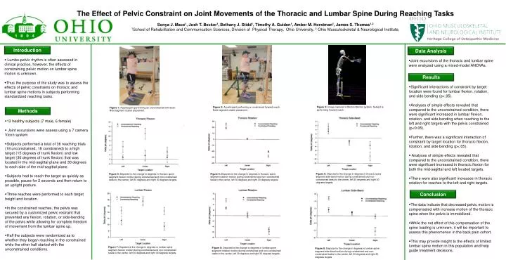
The Effect of Pelvic Constraint on Joint Movements of the Thoracic and Lumbar Spine During Reaching Tasks Sonya J. Mace1, Josh T. Becker1,Bethany J. Stidd1, Timothy A. Guiden1, Amber M. Horstman1, James S. Thomas1,2 1School of Rehabilitation and Communication Sciences, Division of Physical Therapy, Ohio University, 2 Ohio Musculoskeletal & Neurological Institute, Introduction Data Analysis • Lumbo-pelvic rhythm is often assessed in clinical practice, however, the effects of constraining pelvic motion on lumbar spine motion is unknown. • Thus the purpose of the study was to assess the effects of pelvic constraints on thoracic and lumbar spine motions in subjects performing standardized reaching tasks. • Joint excursions of the thoracic and lumbar spine were analyzed using a mixed-model ANOVAs. Results • Significant interactions of constraint by target location were found for lumbar flexion, rotation, and side bending (p<.05). • Analyses of simple effects revealed that compared to the unconstrained condition, there were significant increased in lumbar flexion, rotation, and side bending when reaching to the left and right targets with the pelvis constrained (p<0.05). • Further, there was a significant interaction of constraint by target location for thoracic flexion, rotation, and side bending (p<.05). • Analyses of simple effects revealed that compared to the unconstrained condition, there were significant increased in thoracic flexion for both the mid-sagittal and left located targets. • There were also significant increases in thoracic rotation for reaches to the left and right targets. Figure 3: Image captured in Motion Monitor system. Subject is performing forward reach. Figure 2: A participant performing a constrained forward reach. Note segment cluster placement. Figure 1: A participant performing an unconstrained left reach. Note segment cluster placement. Methods • 13 healthy subjects (7 male, 6 female) • Joint excursions were assess using a 7 camera Vicon system. • Subjects performed a total of 36 reaching trials (18 unconstrained, 18 constrained) to a high target (15 degrees of trunk flexion) and low target (30 degrees of trunk flexion) that was located in the mid-sagittal plane and 30 degrees to each side of the mid-sagittal plane. • Subjects had to reach the target as quickly as possible, pause for 2 seconds and then return to an upright posture. • Three reaches were performed to each target height and location. • In the constrained reaches, the pelvis was secured by a customized pelvic restraint that prevented any flexion, rotation, or side-bending of the pelvis while allowing for complete freedom of movement from the lumbar spine up. • Half the subjects were randomized as to whether they began reaching in the constrained while the other half started with the unconstrained conditions. Figure 6: Depicted is the change in degrees in thoracic spine segment side-bend motion during constrained and non-constrained tasks to the center, left 30 degrees and right 30 degrees targets. Figure 5: Depicted is the change in degrees in thoracic spine segment rotation motion during constrained and non-constrained tasks to the center, left 30 degrees and right 30 degrees targets. Figure 4: Depicted is the change in degrees in thoracic spine segment flexion motion during constrained and non-constrained tasks to the center, left 30 degrees and right 30 degrees targets. Conclusion • The data indicate that decreased pelvic motion is compensated with increase motion of the thoracic spine when the pelvis is immobilized . • While the net effect of this compensation of the spine loading is unknown, it will be important to assess this phenomenon in the back pain cohort. • This may provide insight to the effects of limited lumbar spine motion in this population and help guide treatment decisions. Figure 7: Depicted is the change in degrees in lumbar spine segment flexion motion during constrained and non-constrained tasks to the center, left 30 degrees and right 30 degrees targets. Figure 8: Depicted is the change in degrees in lumbar spine segment rotation motion during constrained and non-constrained tasks to the center, left 30 degrees and right 30 degrees targets. Figure 9: Depicted is the change in degrees in lumbar spine segment side-bend motion during constrained and non-constrained tasks to the center, left 30 degrees and right 30 degrees targets.