Download
1 / 30
300 likes | 383 Views
Cell Structure and Function. Bio 100 Tri-County Tec. College Pendleton, S. C. . Tools to observe cells--. Microscopes light transmission electron scanning electron Each has a unique value to the person studying cells. The Light Microscope. uses visible light for illumination

E N D
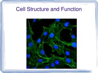
Cell Structure and Function Bio 100 Tri-County Tec. College Pendleton, S. C.
Tools to observe cells-- • Microscopes • light • transmission electron • scanning electron • Each has a unique value to the person studying cells.
The Light Microscope • uses visible light for illumination • magnifies to 1000x (some to 2000x) • good for looking at most cells • living cells can be observed • not good for looking at cell parts
Two important concepts • Magnification defined as “how much larger/bigger the object appears” • Calculated by multiplying the objective by the ocular (40X objective x 10X ocular = 400 X magnification) • Resolution is the ability of a microscope to show two objects as distinct or separate from each other • Catch 22 to be sure • Chalk talk time on resolution
Transmission Electron Micros. • electron beam for illumination • magnifies 100,000x or more • specimen has to be dead and cut into thin sections • good for observing cell parts
Scanning Electron Micros. • electron beam for illumination • can visualize 3D surfaces of whole specimens • specimen must be dead • good for looking a surface architecture of cells
Let’s Review Cell Theory • The cell is the structural and functional unit of life • Whatever “life” is, it begins at the cellular level • All living things are composed of one or more cells • Cells can only come from preexisting cells • Another Catch 22 is spontaneous generation versus biogenesis
The Cell Membrane • Composed of a phospholipid bi-layer with proteins embedded in it • fluid-mosaic model • proteins are randomly distributed in the membrane (mosaic) • proteins are not static in position (fluid) • membrane contains pores
Cell Membrane, ctd. • movement of materials through pores • if smaller than the pores • lipid soluble substances • dissolve in the membrane and move in on the other side • the membrane is selectively permeable • carrier proteins
Organelles of the cell “little organs” that carry out the functions of the cell
Endoplasmic Reticulum • Looks like a maize in the cytoplasm • smooth endoplasmic reticulum (SER) • does not contain ribosomes • rough endoplasmic reticulum (RER) • contains ribosomes • system of channels for internal cellular transport
Golgi Apparatus • Looks like a stack of pancakes • usually near the ER • packages cellular secretions for export from the cell • In some cells, hormones are produced in the ER and the Golgi Apparatus packages these for export.
Ribosomes • Some are located in ER • others float free in cytoplasm • both kinds are the places where proteins are synthesized • composed of RNA (ribosomal-RNA) • protein synthesis takes place in the cytoplasm
Mitochondria • Sites of energy production (ATP synthesis via cellular respiration) • use oxygen to produce ATP • shaped somewhat like a peanut • reactions of energy production take place on numerous membranes that form the inside of the mitochondrion
Centrioles • form the structures that are involved in pulling the chromosomes apart during cell division. • each cell has two • line up at opposite ends of a dividing cell and establish the direction at which division will take place
Nuclear Organelles • located in or associated with the nucleus • nuclear membrane • chromatin • nucleolus • chromosomes • nuclear membrane • controls what enters and exits the nucleus
Nuclear Organelles--2 • Chromatin • stretched out chromosomes • long, thin strands of DNA • Nucleolus • looks like a small nucleus inside the nucleus • synthesizes ribosomes
Nuclear Organelles--3 • chromosomes • before cell division chromatin condenses into chromosomes • composed of DNA • genes are locations on a chromosome that contain directions for making a specific human protein
Cytoskeletal Elements • Microtubules and microfilaments • microtubules • hollow cytoskeletal elements • microfilaments • solid cytoskeletal elements • support the cell from the inside
Organelles of Locomotion • Cilia and flagella • flagella • long, whiplike structures that cause certain cells to move • human sperm cell has a flagellum • cilia • short structures that move materials over the surfaces of certain human tissues
Inclusions • storage areas in the cell • somewhat like closets • surrounded by a membrane • usually spherical in shape like a beach ball • plant cells usually have more inclusions than do animal cells
Lysosomes • Lysosomes • membranous sacs of powerful digestive enzymes • digest worn out cell parts and foreign matter in the cytoplasm • “garbage collectors” of the cell
Peroxisomes • membranous sacs of oxidizing enzymes • detoxify poisons by oxidation • cells produce hydrogen peroxide which is a poison • peroxisomes destroy hydrogen peroxide • “detox” centers
Chloroplasts • Found only in plant cells • location for photosynthesis • production of glucose from carbon dioxide and water • contain chlorophyll (a and b) • Also contain carotenoids and phycobilins
Meanwhile, back at the ranch.. • Plant cells joined together into tissues by cell junctions • Sticky middle lamella cements adjacent cells together • Each plant cell connected to adjoining cells by plasmodesmata • Tiny channels that allow cytoplasm to be continuous between the cells
Extracellular Matrix • Animal cells have elaborate ECM composed mostly of glycoproteins • Tight junctions=continuous belts around cells (membranes of neighboring cells fused at tight junctions) • Anchoring junctions (desmosomes)=rivets fastening cells together into strong sheets • Communicating junctions (gap)=provide cytoplasmic channels between adjacent cells
Eucaryotic vs. Procaryotic One is complex, the other is simple.
Procaryotic Cell • simple, unorganized cell • no membrane-bound organelles • no nuclear membrane • no nucleus • division by binary fission r/t mitosis • small simple ribosomes • examples: bacteria (K. Procaryotae)
Eucaryotic Cell • complex, well organized cell • membrane-bound organelles • nuclear membrane • division involves mitosis • the kind of cell we are composed of and that we have been discussing • members of all other kingdoms
The End The language of the cell is a key to understanding the science of Biology.