Download
1 / 35
370 likes | 567 Views
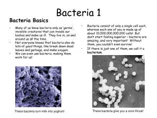
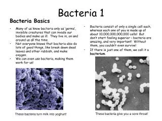
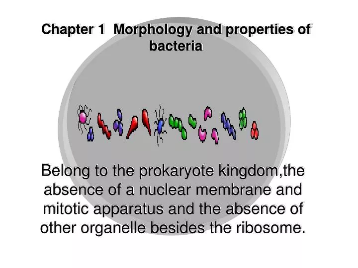
Chapter 1 Morphology and properties of bacteria. Belong to the prokaryote kingdom,the absence of a nuclear membrane and mitotic apparatus and the absence of other organelle besides the ribosome. Section 1 Size and shapes of bacteria. Sizes. The unit of measurement is micrometer (μm).

E N D
Chapter 1 Morphology and properties of bacteria Belong to the prokaryote kingdom,the absence of a nuclear membrane and mitotic apparatus and the absence of other organelle besides the ribosome.
Section 1 Size and shapes of bacteria Sizes The unit of measurement is micrometer (μm)
basic shapes • Spherical: coccus • Straight rod: bacillus • Curved or spiral: vibrio or spirillum • Uncharacteristic (e.g., filamentous, pleomorphic, etc.)
Arrangements • cocci In one plane: Diplococcus Streptococcus In two planes: Tetrad In three planes: Sarcina In random planes:Staphylococcus
Coccus Division in plane (Diplococcl) Neisseria Streptococcus Division in two planes (Chain) (Tetrad) Division in three planes Tetrad (Sarcina) Sarcina Division in Multi- planes Staphylococcus (Clustor) Cell Arrangement
(1)diplococcus S. pneumoniae gonococcus
(2)streptococcus α-hemolytic streptococcus
(3)staphylococcus S. aureus
Arrangements • Bacilli Only in one plane Bacillus Streptobacillus Coccobacillus
Arrangements • Spiral bacteria in single cells
Section 2 Structures of Bacterial cells • Basic Cell wall Cellular membrane Cytoplasm nucleoid • Special Capsule Flagellum Pilus Spore
Cell wall-Components • Peptidoglycan Backbone: alternating N-acetylmuramic acid (NAM) and N-acetylglucosamine (NAG) A set of identical tetrapeptide side chains A set of identical pentapeptide cross bridges(G+)
peptidoglycan N-acetylglucosamine N-acetylmuramic acid Lysozyme breaks this bond L-ala D-glu L-lys D-ala D-ala L-ala D-glu m-DAP D-ala D-ala penicillin pentaglycine bridge Staphylococcus aureus 金黄色葡萄球菌 E.coli 大肠杆菌
Cell wall-Components • Teichoic acid: Organisms: G+bacteria Types: the wall teichoic acid the membrane teichoic acid Function: acts as a specific antigenic determinant Relation to pathogenicity
Wall teichoic acid (LTA)
Cell wall-Components • Outer membrane Organisms: G-bacteria Composition: Lipoprotein Lipid bilayer Lipopolysaccharide (LPS): lipid A core polysaccharide O-polysaccharide
Outer membrane Functions: prevents toxic materials from entering functions as a harmful endotoxin possesses the property of antigenic specificity
GlN-Mur GlN-Mur GlN-Mur GlN-Mur GlN-Mur GlN-Mur GlN-Mur GlN-Mur GlN-Mur Gln-Mur Gln-Mur Gln-Mur GlN-Mur GlN-Mur GlN-Mur GlN-Mur GlN-Mur GlN-Mur GlN-Mur GlN-Mur GlN-Mur GlN-Mur GlN-Mur GlN-Mur GlN-Mur GlN-Mur GlN-Mur GlN-Mur GRAM POSITIVE Lipoteichoic acid Peptidoglycan-teichoic acid Cytoplasmic membrane Cytoplasm GRAM NEGATIVE Lipopolysaccharide Porin Outer Membrane Braun lipoprotein Periplasmic space Inner (cytoplasmic) membrane Cytoplasm
Properties of the cell walls of Gram-positive and Gram-negative bacteria
Cell wall Functions • gives the bacterium its shape • Protecting the cell • Constituting major surface antigens • Permitting the free diffusion of low-molecular-weight hydrophilic compounds • Having pathogenic material Gram-positive Gram-negative
The mechanism of some antibiotics and enzymes killing bacteria • penicillin: Binds to transpeptidase and blocks the formation of whole cell wall of Gram-positive bacteria • Lysozyme: Destroys the peptidoglycan by cleaving the β-1,4 glycosidic bonds between NAM and NAG G+ G-
G+ and G- N-acetylglucosamine N-acetylmuramic acid Lysozyme breaks this bond L-ala D-glu L-lys D-ala D-ala L-ala D-glu m-DAP D-ala D-ala penicillin pentaglycine bridge Staphylococcus aureus E.coli
Penicillin and cephalosporin • Results of enzyme digestion:Protoplast Spheroplast
L forms • Definition: bacteria that their peptidoglycan is destroyed or lost by various factors but they can survive under highly osmotic environment. • Types: Protoplasts(G+) and spheroplasts (G-) • Shape: polymorphism • Staining property: gram-negative • Grow environment:high osmotic • Pathogenicity: chronic
A.Colony of normal bacteria B.fried egg colony of L-form bacteria C.Granular colony of L-form bacteria D.filamentous colony of L-form bacteria
L forms are able to grow and divide • Caused by antibiotic • Some L forms can revert to the normal form upon removal of the inducing stimulus
producing chronic infections,being resistant to antibiotic treatment.
B.Cell membrane(Cytoplasmic membrane) • Composition: composed primarily of proteins and lipids. • It is a thin,elastic cytoplasmic membrane,which is 5~10nm thick • Visible in some ultrathin sections • Examined with the electron microscope
Function active transport respiratory chain component biosynthesis excretion