Download
1 / 44
440 likes | 483 Views
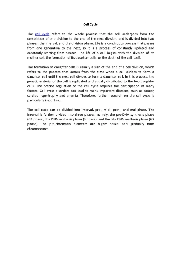
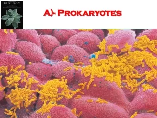
Cell division in prokaryotes takes place in two stages The DNA is replicated The cell elongates, then splits into two daughter cells The process is called binary fission. 7.1 Prokaryotes Have a Simple Cell Cycle. Fig. 7.1 Cell division in prokaryotes.

E N D
Cell division in prokaryotes takes place in two stages The DNA is replicated The cell elongates, then splits into two daughter cells The process is called binary fission 7.1 Prokaryotes Have aSimple Cell Cycle
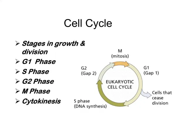
Cell division in eukaryotes is more complex than in prokaryotes because 1. Eukaryotic contain far more DNA and a nucleus 2. Eukaryotic DNA is packaged differently It is in linear chromosomes compacted with proteins 7.2 Eukaryotes Have aComplex Cell Cycle
Eukaryotic cells divide in one of two ways Mitosis Occurs in somatic (non-reproductive) cells Meiosis Occurs in germ (reproductive) cells Results in the production of gametes 7.2 Eukaryotes Have aComplex Cell Cycle
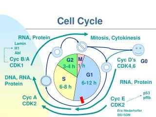
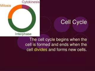
The complex cell cycle of eukaryotic cell is composed of several stages Interphase • G1 phase • Primary growth phase • S phase • DNA replication • G2 phase • Microtubule synthesis, growth, mitochondria replication • M phase • Chromosomes pull apart • C phase • Cytoplasm divides Mitosis Cytokinesis
7.3 Chromosomes • Chromosomes were first observed by the German embryologist Walther Fleming in 1882 • The number of chromosomes varies enormously from species to species • The Australian ant Myrmecia spp. has only 1 pair • Some ferns have more than 500 pairs • Chromosomes exist in somatic cells as pairs • Homologous chromosomes or homologues
Fig. 7.3 • Replicated chromosomes consist of two sister chromatids • These are held together at the centromere • Diploid cells have two copies of each chromosomes
Fig. 7.4 7.3 Chromosomes • Humans have 46 chromosomes The 23 pairs of homologous chromosomes can be organized by size This display is termed a karyotype
7.3 Chromosomes • Chromosomes are composed of chromatin • Complex of DNA (~ 40%) and proteins (~ 60%) • A typical human chromosome contains about 140 million nucleotides in its DNA • This is equivalent to • About 5 cm in stretched length • 2,000 printed books of 1,000 pages each! • In the cell, however, the DNA is coiled
Interphase (“in-between divisions”) • Cell grows (G1), replicates DNA (S), and grows some more (G2) • Mitosis • Prophase (“prepares”) • Nuclear envelope breaks down • Chromosomes condense further • Spindle apparatus is formed • Metaphase (“middle”) • Chromosomes align along the equatorial plane • Spindle fibers attach to the chromosomes
Anaphase (“away”) • Sister chromatids separate • They are drawn to opposite poles by shortening of the microtubules attached to them • Telophase (“tears into two”) • Nuclear envelope reappears • Chromosomes decondense • Spindle apparatus is disassembled • Cytokinesis • Two diploid daughter cells form • Mitosis
Fig. 7.8 Animal cells • Cytokinesis • Cleavage furrowforms, pinching the cell in two Plant cells • Cell plateforms, dividing the cell in two
Fig. 7.10 7.5 Controlling the Cell Cycle • The eukaryotic cell cycle is controlled by feedback at three checkpoints
Fig. 7.13 7.6 What is Cancer? • Cancer is unrestrained cell growth and division • The result is a cluster of cells termed a tumor • Benign tumors • Encapsulated and noninvasive • Malignant tumors • Not encapsulated and invasive • Can undergo metastasis • Leave the tumor and spread throughout the body
7.6 What is Cancer? • Most cancers result from mutations in growth-regulating genes • There are two general classes of these genes • 1. Proto-oncogenes • Encode proteins that simulate cell division • If mutated, they become oncogenes • 2. Tumor-suppressor genes • Encode proteins that inhibit cell division • Cancer can be caused by chemicals, radiation or even some viruses
The p53gene plays a key role in the G1 checkpoint of cell division The p53 protein (the gene’s product), monitors the integrity of DNA If DNA is damaged, the protein halts cell division and stimulates repair enzymes If the p53 gene is mutated Cancerous cells repeatedly divide No stopping at the G1 checkpoint 7.7 Cancer and Controlof the Cell Cycle
7.8 Curing Cancer • Potential cancer therapies are being developed to target seven different stages in the cancer process • Stages 1-6 • Prevent the start of cancer within cells • Focus on the decision-making process to divide • Stage 7 • Act outside cancer cells • Prevents tumors from growing and spreading
Fig. 7.15 New molecular therapies for cancer Stopping tumor growth Receiving the signal to divide Stepping on the gas Passing the signal via a relay switch Amplifying the signal Releasing the “brake” Checking that everything is ready
7.9 Discovery of Meiosis • Meiosis was first observed by the Belgian cytologist Pierre-Joseph van Beneden in 1887 • Gametes (eggs and sperm) contain half the complement of chromosomes found in other cells • The fusion of gametes is called fertilization or syngamy • It creates the zygote, which contains two copies of each chromosome
Fig. 7.16 Contain one set of chromosomes • Sexual reproduction • Involves the alternation of meiosis and fertilization • Asexual reproduction • Does not involve fertilization Contains two sets of chromosomes
7.10 The Sexual Life Cycle • The life cycles of all sexually-reproducing organisms follows the same basic pattern • Haploid cells or organisms alternate with diploid cells or organisms • There are three basic types of sexual life cycles
7.11 The Stages of Meiosis • Meiosis consists of two successive divisions, but only one DNA replication • Meiosis I • Separates the two versions of each chromosome • Meiosis II • Separates the two sister chromatids of each chromosome • Meiosis halves the number of chromosomes
Haploid gametes PROPHASE I Germ-line cell II TELOPHASE METAPHASE I II II I ANAPHASE II I Fig. 7.22 How meiosis works Diploid cell
Meiosis I • Prophase I • Homologous chromosomes pair up and exchange segments • Metaphase I • Homologous chromosome pairs align at random in the equatorial plane • Anaphase I • Homologous chromosomes separate and move to opposite poles • Telophase I • Individual chromosomes gather together at each of the two poles
Fig. 7.20 • The longest and most complex stage of meiosis • Homologous chromosomes undergo synapsis • Pair up along their lengths • Meiosis I • Prophase I • Crossing overoccurs
Interkinesis Fig. 7.23
Meiosis II • After meiosis I there is a brief interphase • No DNA synthesis occurs • Meiosis II is similar to mitosis, but with two main differences • 1. Haploid set of chromosomes • 2. Sister chromatids are not identical
Prophase II • Brief and simple, unlike prophase I • Metaphase II • Spindle fibers bind to both sides of the centromere • Anaphase II • Spindle fibers contract, splitting the centromeres • Sister chromatids move to opposite poles • Telophase I • Nuclear envelope reforms around four sets of daughter chromosomes • Meiosis II
No two cells are alike Fig. 7.23
7.12 Comparing Meiosis and Mitosis • Meiosis and mitosis have much in common • However, meiosis has two unique features • 1. Synapsis • Homologous chromosomes pair all along their lengths in meiosis I • 2. Reduction division • There is no chromosome duplication between the two meiotic divisions • This produces haploid gametes
Sexual reproduction increases genetic diversity through three key mechanisms 1. Independent assortment 2. Crossing over 3. Random fertilization 7.13 Evolutionary Consequencesof Sex
Fig. 7.26 Independent assortment • In humans, a gamete receives one homologue of each of the 23 chromosomes • Humans have 23 pairs of chromosomes • 223 combinations in an egg or sperm • 8,388,608 possible kinds of gametes Three chromosome pairs 23 combinations
Fig. 7.20 Crossing over • DNA exchanges between maternal and paternal chromatid pairs This adds even more recombination to independent assortment that occurs later
Random fertilization • The zygote is formed by the union of two independently-produced gametes • Therefore, the possible combinations in an offspring • 8,388,608 X 8,388,608 = • 70,368,744,177,664 • More than 70 trillion! • And this number does not count crossing-over
Importance of Generating Diversity • Genetic diversity is the raw material that fuels evolution • And no genetic process generates diversity more quickly than sexual reproduction