Download
1 / 68
680 likes | 681 Views
Learn about the male reproductive system, including the functions of the testes, spermatozoa production, and the accessory organs involved in reproduction. Discover the pathway of sperm cells and the different secretions that make up semen.

E N D
Memmler’s The Human Body in Health and Disease11th edition Chapter 23 The Male and Female Reproductive Systems
Reproduction Asexual (nonsexual) • Divide by themselves • Produce genetically identical offspring. Sexual • Produce genetically similar offspring. • Specialized sex cells (germ cells or gametes) • Male are spermatozoa (sperm cells) • Female are ova (egg cells)
Meiosis Cell division process • Halves the number of chromosomes in cell from 46 to 23 • Produces gametes or germ cells • Human gametes have 23 chromosomes • In males, occurs from puberty until death. • In females, begins in womb and is completed each month at ovulation.
Checkpoint 23-1:What is the process of cell division that halves the chromosome number in a cell to produce a gamete?
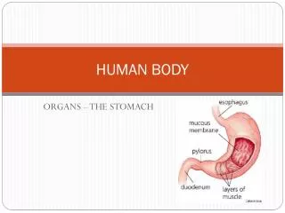
The Male Reproductive System Two groups of organs • Primary • Gonads – sex gland – produce the germ cells and manufacture hormones • Testes • Accessory • Ducts • Exocrine glands
The Testes • Testes or testicles • Located outside the body proper (cooler temperature than body) and are suspended in a sac between the thighs called the scrotum • Two functions: production of sperm and secretion of male hormone, testosterone • Testes normally descend into the scrotum during the last 2 months of fetal development. Failure of testes to descend into the scrotum is called cryptorchidism and can result in sterility.
Internal Structure Specialized tissue in testis • Seminiferous tubules form sperm • Sustentacular cells – nourish and protect developing spermatozoa. • Interstitial cells produce the male hormones called androgens. Testosterone is an androgen.
Testosterone Male sex hormone functions • Develops and maintains reproductive structures • Develops spermatozoa • Develops secondary sex characteristics • Deeper voice • Broader shoulders • Narrower hips • More muscle tissue • More body hair
The Spermatozoa Individual cells manufactured in seminiferous tubules • Head • Nucleus (no cytoplasm) • Acrosome contains enzymes that help the sperm cell to penetrate the ovum • Midpiece • Mitochondria provide energy for movement • Tail (flagellum) propels the sperm through the femal reproductive system to the ovum Figure 23-4 Page 488
Question:What is the name of the cells that nourish and protect developing spermatozoa?a. sustentacular cells b. interstitial cellsc. testicular cells
Checkpoint 23-2:What is the male gonad?What is the main male sex hormone?Checkpoint 23-3:What is the male sex cell (gamete) called?Checkpoint 23-4:What are the main subdivisions of a spermatozoon?
Accessory Organs System of ducts that transports spermatozoa • Seminferous Tubules – as sperm form they gather here and move into a series of genital ducts where they mature • Epididymis – first part of duct system. It is where sperm mature, become motile and fertile. The walls of the epididymis contract and push the sperm into the vas deferens. • Ductus deferens (vas deferens) –a coiled duct that conveys sperm from the epididymis to the ejaculatory duct and the urethra. • Seminal vesicle twisted muscular tubes with many small outpouchings found behind the urinary bladder. They They secrete a sugar and other substances that provide nourishment for the perm. The seminal fluid makes up about 60% of the semen’s volume. • Ejaculatory duct is formed by the union of the vas deferns with the seminal vesicle.
Sperm Cell PathwaySeminferous Tubules (Germinal epithelium of testis produces sperm)Epididymis (temporary storage of sperm cell)Vas Deferens (Transports sperm after ejaculation)Ejaculatory DuctUrethra Through the PenisUrethral Orifice
Semen Various secretions are added to sperm as they travel through the genital ducts. The secretions come from 3 glands: • Seminal vesicles – secretes a thick material rich in sugar, vitamin C and prostaglandins (fats). • Prostate gland – secretes an alkaline substance that plays a role in increasing sperm motility and counteracting the acidic environment of the vagina thereby protecting the sperm as they enter the woman’s body. During ejaculation the smooth muscle of the prostate gland contracts and forces the secretions into the urethra.
Bulbourethral glands (Cowper glands) a pair of tiny glands that secrete thick mucus into the urethra. The mucus serves as a lubricant during sexual intercourse. • Functions: • Nourish spermatozoa • Transport spermatozoa • Neutralize acidity of male urethra and female vagina • Lubricate reproductive tract during intercourse • Prevent infection with antibacterial enzymes and antibodies
Checkpoint 23-6:What glands, aside from the testis, contribute secretions to semen?
The Urethra and Penis Urethra • Carries urine from bladder • Carries reproductive cells outside body Penis carries urine through the urethra to the outside of the body and acts as the organ of sexual intercourse (copulation) by depositing sperm in the female reproductive tract. • Glans penis – enlarged tip of penis. • Prepuce (foreskin)- a loose fold of skin that covers the glans penis and is removed in a circumcision. • Penis and scrotum together make up male external genitalia.
Ejaculation Reflex centers in spinal cord initiate process • Smooth muscle contraction in prostate • Skeletal muscle contraction in pelvic floor • Forceful expulsion of semen through urethra to outside • Hypothalamus (conscious control) • Parasympathetic nerves • Viagra- promotes vasodilation • Control of emission and ejaculation • sympathetic nerves- muscle contraction
Hormonal Control of Male Reproduction Anterior pituitary produces hormones that control testes • Follicle-stimulating hormone (FSH) • Promotes spermatozoa formation • Luteinizing hormone (LH) • Stimulates testosterone development • Aids sperm cell development • Hypothalamus secretes hormones that trigger the release of FSH & LH. Activity of hypothalamus is regulated by a negative feedback mechanism. When blood level of testosterone increases, hypothalamus secretes less releasing hormone. When level of testosterone decreases, hypothalamus secretes more releasing hormone.
Checkpoint 23-7:What two pituitary hormones regulate both male and female reproduction?
Question:What is the anatomic name for the foreskin of the penis?a. acrosome b. corpus cavernosum c. prepuce
The Effects of Aging on Male Reproduction Decreasing • Testosterone production • Spermatozoa production • Prostate secretions • Seminal vesicle secretions
Disorders of the Male Reproductive System • Infertility – a lower than normal ability to reproduce • Sterility complete inability to reproduce • Oligospermia - Low sperm count. • Tubule damage of the testes can be caused by X-rays, Infections, Toxins, Malnutrition • Vasectomy – intentional sterilization
Structural Disorders • Cryptorchidism – failure of testis to descend into scrotum. Surgical correction is required if the testis does not descend by 1 year of age otherwise sterility will result. • Torsion of testis – twisting of spermatic cord resulting from a rotation of the testis. This requires emergency surgery to correct and may involve removal of testis – orchiectomy. • Hernia – during development the testis pushes its way through the muscles and connective tissues of the abdominal wall carrying with it the blood vessels and other structures that form the spermatic cord. • Phimosis – a tightness of the prepuce (foreskin) that can be remedied by circumcision.
Infections • Sexually transmitted infection (STI) spread through sexual contact. Most common involve: Chlamydia and Gonorrhea. • Genital herpes – caused by a virus and characterized by blisters on and around genital organs • Syphilis – is caused by spirochete (Treponema pallidum) that causes genital ulcers. • Epididymitis - is swelling (inflammation) of the epididymis caused by organisms from an STI or urinary tract infection that traveled through the ducts of the reproductive system to the epididymis. • Prostatitis – prostatic inflammation usually caused by bacterial infection especially E. coli • Orchitis – inflammation of the testis
Checkpoint 23-8:What are some infectious diseases of the reproductive tract?
Tumors • Prostate tumors • Can be benign or malignant • Most common cancer of males in U.S. • Testicular tumors affect young to middle-aged adults. These tumors arise in germ cells and can metastasize through the lymphatic system . Early detection and regular testicular self-examination (TSE) improves the chances for effective treatment.
The Female Reproductive System • Ovaries (paired female gonads) – where female sex cells or ova are formed • Uterus – the organ that holds and nourishes a developing infant • Passageways • Oviduct (fallopian tube or uterine tube) • External genital organs
Figure 23-8 Page 493 Female reproductive system. The enlargement (right) shows ovulation. Zooming In: What is the deepest part of the uterus called?
The Ovaries • Located in pelvic abdomen • Small, flattened oval body measuring about 1.6 inches in length, .8 inches in width and .4 inches in depth • Held in place by ligaments that attach them to uterus and body wall
The Ova and Ovulation • Ovarian follicle (graafian follicle) • Holds ripening ova • Each month during the reproductive years several ripen but usually one is released • Secretes estrogen – stimulates the growth of the uterine lining • Ruptures to discharge egg cell (ovulation) when it has ripened. • Oviduct – after ovulation the egg cell is swept into this tube that leads to uterus
Checkpoint 23-9:What is the female gonad called?Checkpoint 23-10:What is the female gamete called?
Question:What is the name of the passageway the ovum travels through to get to the uterus?a. oviductb. cervical canalc. ovarian ligament
Answer:a. oviduct (also known as the fallopian tubeor uterine tube)
Formed from remains of follicle after ovum is expelled Secretes estrogen and progesterone If fertilization does NOT occur it deteriorates in about 10 days and then is called the corpus albicans (white body) which cannot secrete hormones Remains active during pregnancy Sometimes the corpus luteum fills with fluid & forms an ovarian cyst. A blood-filled cyst is called a chocolate cyst. These cysts can resolve on their own but may require surgery. The Corpus Luteum
Checkpoint 23-11:What is the structure that surrounds the egg as it ripens?Checkpoint 23-12:What is the process of releasing an egg cell from the ovary called?
In females Oviducts – (fallopian tubes, uterine tubes) transport ova. They extend from either side of the uterus to the ovaries. The funnel-shaped end is called the infundibulum & has fingerlike projections called fimbriae. Ova cannot move on their own. Fimbriae sweep ova from the ovary into the oviducts. The egg then moves slowly toward the uterus by peristaltic activity of oviducts. Oviducts are the usual site of fertilization of the egg by the sperm. The journey through the oviducts takes about 4 to 5 days. Accessory Organs
Accessory Organs continued • Uterus – (womb) where the fetus develops to maturity. • Vagina – muscular tube that extends from the cervix to the vaginal opening in the perineum. The opening is usually covered by a thin membrane called the hymen. • Greater vestibular glands – (Bartholin) two mucus-producing glands that secrete into an area near the vaginal opening known as the vestibule. These glands provide lubrication during intercourse. • Vulva and perineum – external genitalia • Vulva has 2 pairs of lips or labia, the clitoris (an organ of sensitivity) and related structure. • Perineum - The area between the anus and the vulva in the female.
The Oviducts • Extend from near ovary to uterus • Not connected to ovary • Fimbriae produce current that sweeps ova into oviduct • Cilia in tube lining and peristalsis of tube move ova
Checkpoint 23-13:What does the follicle become after ovulation?
The Uterus • Organ where fetus develops to maturity • Corpus • Body is the wider upper region • Cervix • Lower narrow region that opens into vagina • Fundus – the upper dome-shaped region above the entrance of the oviducts • Broad ligaments support the uterus. They extend from each side to the lateral body wall. • Uterus has 3 Parts: • Myometrium - a middle, smooth muscular layer • Endometrium – an inner layer uterine lining that has 2 layers: the basilar layer & the functional layer. The basilar layer is thin and vascular & lies next to myometrium. The functional layer responds to ovarian hormones & thickens in preparation for fertilized egg. It is the layer that sloughs off during menstruation when fertilization has not occurred. • Epimetrium – outer layer.
The Vagina Distal part of birth canal that opens to outside of body • Fornix – a circular recess formed when the cervix dips into the superior portion of the vagina • Posterior fornix – the deepest area of the fornix, located behind the cervix. • Cul-de-sac (rectouterine pouch or pouch of Douglas) An extension of the peritoneal cavity between the rectum and back wall of the uterus. • Hymen • Greater vestibular (Bartholin) glands -
Figure 23-11 Page 495 Female reproductive system (sagittal section). This view shows the relationship of the reproductive organs to each other and to other structures in the pelvic cavity. Zooming In: Which has the more anterior opening, the vagina or the urethra?
The Menstrual Cycle • Controlled by pituitary hormones regulated by hypothalamus • Cyclic pattern • Regulated by hormonal feedback • Averages 28 days • The first day of menstrual is consider the first day of the cycle
Beginning of the Cycle • Several follicles, each containing an ovum begin to develop in the ovary but only one will release an ovum per month • Follicle produces increased amounts of estrogen as it matures. Estrogen is carried in the bloodstream to the uterus where it starts preparing the endometrium for a possible pregnancy. • Thickens endometrium • Elongates uterine secretion glands • Inhibits release of FSH • Stimulates pituitary to release LH (Luteinizing hormone)
Ovulation • In an average 28 day cycle, ovulation occurs on day 14 • LH surge in blood occurs about 1 day before ovulation • LH causes ovulation • Transforms ruptured follicle into corpus luteum that produces estrogen and progesterone • Endometrium thickens due to influence of estrogen and progesterone. Also, glands and blood vessels increase in size. • FSH and LH are inhibited due to a negative feedback mechanism