Download
1 / 48
480 likes | 499 Views
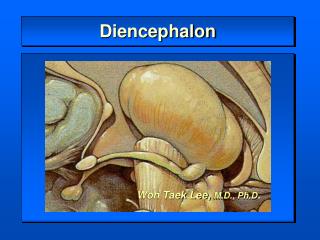
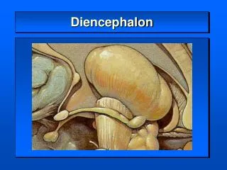
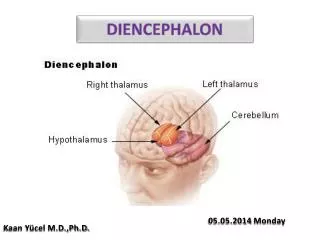
Microscopy of the diencephalon. András Csillag In collaboration with Prof. Á. Dobolyi and Dr. V. Vereczki Semmelweis University, Department of Anatomy , Histology and Embryology. The position of the diencephalon in the brain. Parts of the diencephalon. Thalamus Epithalamus

E N D
Microscopy of the diencephalon András Csillag In collaborationwith Prof. Á. Dobolyi and Dr. V. Vereczki Semmelweis University, Department of Anatomy, Histology and Embryology
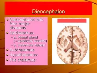
Parts of the diencephalon • Thalamus • Epithalamus • Pineal body • Habenula • Trigonum habenulae • Habenular nuclei • Stria medullaris • Habenular commissure • Metathalamus • Medial geniculate body • Lateral geniculate body • Subthalamus • Subthalamic nucleus • Zona incerta • H fields of Forel • Hypothalamus
Thalamic subdivisions of human brain Williams et al., (1989) Gray’s Anatomy, Churchill Livingstone
Functional groups of thalamic nuclei Mammaliansystem Nucl. ventralis post. VPL/VPM SENSORY THALAMUS MOTOR THALAMUS Ventral tier nuclei (VA/VL) Anterior nuclei VISCERAL AND LIMBIC THALAMUS Intralaminar nuclei Midline nuclei Mediodorsal nucleus
Frontal sections of the thalamus Anterior section Middle section
Functional classification of thalamic nuclei • Specific nuclei: specific input, project to specific part of the cortex • - sensory relay nuclei: VPL, VPM, CGL, CGM • - motor relay nuclei: VA, VL • - limbic relay nuclei: AV, AD, AM • Associate nuclei: cortical input, project to associate areas of the cortex • MD, LD, LP, pulvinar • Non-specific nuclei: ascending input, diffuse projection to the cortex • midline and intralaminar nuclei • Nuclei not projecting to the cerebral cortex • n. reticularis thalami, n. parafascicularis, n. subparafascicularis
Cortical projections of (specific and associate) thalamic nuclei mediosagittal view lateral view
The VPL relays sensory inputs from the body to the cerebral cortex
The VPM relays sensory inputs from the head to the cerebral cortex
Relay of gustatory inputs to the cortex takes place in the VPMpc
Afferent and efferentprojections of the motor relaynuclei in theventralcolumn of thelateralnucleargroup (‚ventraltier’ nuclei) Specific thalamic motor relay nuclei: Ventral anterior nucleus (VA) Afferents (inputs): basal ganglia (pallidum) Efferents (projections): premotor and supplementary motor cortex Ventral lateral nucleus (VL) Afferents (inputs): cerebellum (dentate nucleus) Efferents (projections): primary motor cortex (precentral gyrus )
Neuronal circuit of information relay in the specific thalamic nuclei
The IMMC Complex 1. Midline thalamic nuclei: Paraventricular Parataenial Intermediodorsal Rhomboid Reuniens 2. Mediodorsal nucleus 3. Intralaminar nuclei: Rostral group Central medial Paracentral Central lateral Caudal group Centre médian Parafascicular
Midline thalamic group: paraventricular nucleus Input from preoptic area Output to medial orbitofrontal cortex Output to dorsomedial hypothalamus (DMH) Input from (output to ?) midbrain periaqueductal gray (PAG)
Mediodorsal thalamic nucleus Output to prefrontal cortex and anterior cingulate cortex Output to amygdala Reciprocal (?) connections with lateral hypothalamus and PAG
Intralaminar thalamic group Output to non-limbic (visceral, sensory and motor) cortices Output to (mainly) dorsal striatum Output to hypothalamus ? Output to reticular thalamic nuclei Nissl-stainedmonkeybrainatthelevel of thecaudalintralaminarnuclei
Major inputs and projections of midline and intralaminar thalamic nuclei Spino-reticulo-thalamic tract – part of the „ascending reticular activating system”, a regulator of cortical alertness
PULV. CGL VL DM LP LD CGM VA VPL+VPM Cortical efferents of thalamic nuclei
Thalamic reticular nucleus: a regulator of the relay function of specific thalamic nuclei
The hypothalamus • Thalamus • Epithalamus • Pineal body • Habenula • Trigonum habenulae • Habenular nuclei • Stria medullaris • Habenular commissure • Metathalamus • Medial geniculate body • Lateral geniculate body • Subthalamus • Subthalamic nucleus • Zona incerta • H fields of Forel • Hypothalamus
Essentialdifferencesbetweenthestructure and functions of the thalamus and hypothalamus Thalamus:- welldelineatednuclei- relay and modulation of corticalinputs • Hypothalamus: • Theneuronswithdifferentfunctionsareintermingled, notclearlyseparated • Homeostaticregulatoryfunctionsdonotrequirecorticalprocessing
Tuberal and posteriorhypothalamicregions Tuberal hypothalamic region Posterior hypothalamic region
Hypothalamo-spinal tract and other descending pathways regulating vegetative functions
Regulatory functions of hypothalamic nuclei • Vegetative regulations • Neuroendocrine regulations • Salt and water balance • Food intake and body weight • Temperature • Circadian rhythms • Sleep • Reproduction
Neuralelements of homeostaticregulations NTS: nucleus of the solitary tract, PBN: parabrachial nucleus, RF: reticular formation, DMX: dorsal motor vagus nucleus, ILN: intermediolateral column (nucleus) of the spinal cord
Circadial regulation of neural functionsPACEMAKER: SUPRACHIASMATIC NUCLEUS (SCN)
5. control of circadian rhytms: retinohypothalamic tract Medial zone Suprachiasmaticus nucleus (VIP):retinohypothalamic tract of rat (Scale: 100 µm. OX= opticchiasm;SCH=suprachiasmatic nucleus)
SUBTHALAMUS - Ventralfromthalamus - Caudal and lateralfromhypothalamus - Prosomere 3 + Peduncularhypo- thalamus (basal part) Main parts: Subthalamicnucleus (Luys) Zonaincerta Fields of Forel
EPITHALAMUS Habenular nuclei Septohabenular pathway (stria medullaris thalami) Retroflex fascicle (fasciculus retroflexus) – habenulo-interpeduncular pathway Retroflex fascicle – border between prosomers 1 and 2 Part of the limbic system, From stress evasion to value-based decision making, Connections with the basal ganglia and the serotonergic system of brainstem (raphe nuclei)