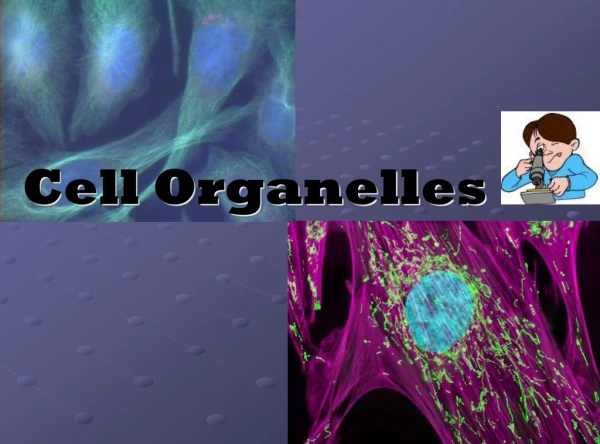
Download
1 / 32
320 likes | 487 Views
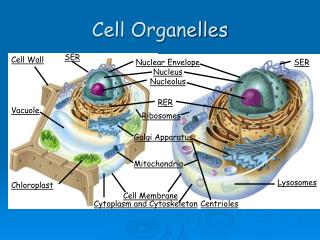
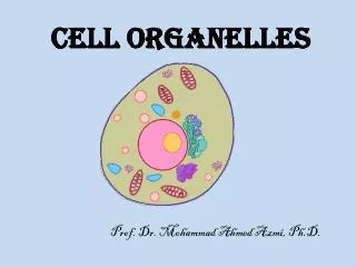
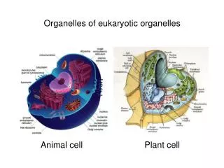
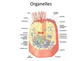
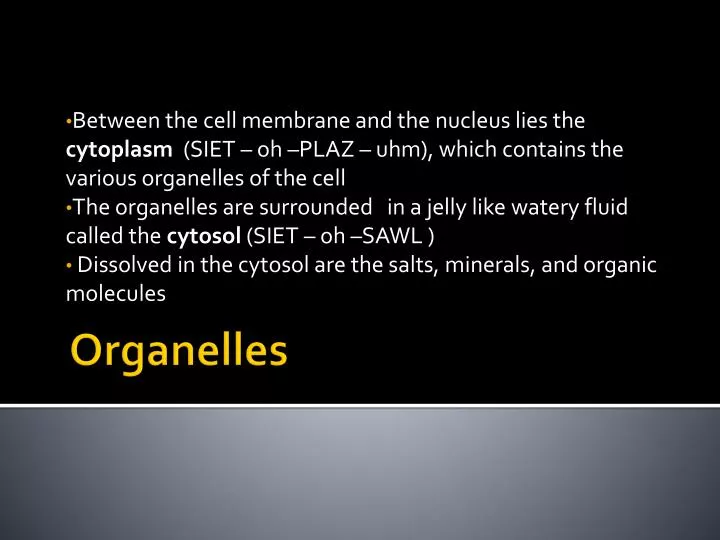
Between the cell membrane and the nucleus lies the cytoplasm (SIET – oh –PLAZ – uhm), which contains the various organelles of the cell The organelles are surrounded in a jelly like watery fluid called the cytosol (SIET – oh –SAWL )

E N D
Between the cell membrane and the nucleus lies the cytoplasm (SIET – oh –PLAZ – uhm), which contains the various organelles of the cell The organelles are surrounded in a jelly like watery fluid called the cytosol (SIET – oh –SAWL ) Dissolved in the cytosol are the salts, minerals, and organic molecules Organelles
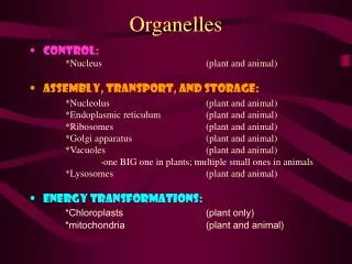
Mitochondria • Comes from the Greek mitos, meaning “thread”, and chondrion, meaning “grain” • Are the power houses of the cell and contain the molecular machinery for the conversion of energy from the breakdown of glucose into adenosine triphosphate (ATP), the energy used by all cells. • (MIET – oh –KAHN –dree –uh)
Mitochondria • The energy stored in the high energy phosphate bonds of ATP is then available to power cellular functions. • Mitochondria are mostly protein, but some lipid, DNA and RNA are present. • The unique structure of these organelles can be seen under the electron microscope.
DIAGRAM • The metabolic activity of the cell is related to the number of cristae (KRIS-tee) and the number of mitochondria within a cell. • Cells with large amounts of metabolic activity, such as heart muscle/sperm cells, have many well developed mitochondria.
These generally spherical organelles have an outer membrane surrounding an inner membrane that folds (cristae) to provide more space for chemical reactions • These reactions are oxidative phosphorylation and electron transport enzymes. • Those big words are just ways that ATP is formed
DIAGRAM • Mitochondria have their own DNA, and new Mitochondria are created only when existing ones grow and divide.
RIBOSOMES (RIE-buh-SOHMZ) • Ribosomes are small organelles composed of ribosomal RNA (rRNA) and 80 some different proteins. • Inside the cell’s nucleus rRNA and proteins are cobimed to form Ribosomes • The Ribosomes are then transported to cytosol • Ribosomes play an important part in the production of proteins.
Ribosome's are the most common organelle in many cells • Ribosome’s do not have an outer membrane
Endoplasmic Reticulum (ER)(EN-doh-PLAZ-mikri-TIK-yuh-luhm) • A system of membrane tubes and sacs • The ER functions acts like a highway for the cell, providing a road for molecules to move form one cell to the other. • The number of ER in a cell depends on the activity of the cell and can change
Two types of ER • Rough Endoplasmic Reticulum • - (Rough ER) • - has attached ribosome's that give it a rough appearance • Is usually in cells that make a large to be exported from or imported to the cell
Smooth Endoplasmic reticulum • Smooth ER • -is not covered with ribosomes, giving it a smooth appearance • - Smooth ER is involved with the production of steroids in gland cells, the control of calcium levels in muscle cells, and the breakdown of toxic substances by liver cells.
Golgi Complex (GOHL – jee) • The Golgi Complex is responsible for the packaging of proteins in the cell. • The Golgi is a curved membrane stack resembling a stack of pancakes. • The Golgi bodies package proteins produced in the ribosome and transport them via the ER. • Proteins within the rough ER bud off and are transported to the Golgi where they are further modified and packaged for export.
All this packaging is necessary so the cell is able to recognize the protein as 'self'. • If the protein is not recognized the immune system will treat the protein as a foreign body known as an antigen. • When this happens the immune system undergoes an immune response. • Viruses and bacteria that cause disease are recognized as antigens(foreign proteins) and attacked immediately.
In this graphic a protein is produced in a ribosome on the ER and transported to the golgi complex where it is packaged and exported.
Lysosomes(LIE-suh-sohmz) • Are small, spherical organelles that enclose more than 40 hydrolytic enzymes • These enzymes can digest proteins, carbohydrates, lipids, DNA and RNA. • They may also digest old organelles and viruses and bacteria that has invaded a cell.
Lysosomes are common in the cells of animals, fungi, and protists, but they are rare in plant cells. • For example, the human hand begins as a solid structure in the embryo. • As the embryo develops, lysosomes enzymes selectively destroys tissue to form the spaces the spaces between the fingers.
Cytolskeleton • Just as your body needs a skeleton to keeps its size and shape, a cell need a structure to maintain its shape and size. • This is called a Cytoskeleton, a network of long protein strands located in the cytosol. • In addition to providing support, the cytoskeleton helps with the movement of organelles with in the cytosol.
Microfilaments and Microtubules • Two major components of the cytolskeleton are microfilaments and microtubules. • Microfilaments are threads made of a protein called actin. • Each microfilament is made of many actins molecules that are linked together to form a polymer chain. • The microfilaments help with cell movement and play a role in the contraction of muscles.
Microtubules • Hollow tubes that extend outward from a central point near the nucleus to various cites near the cell membrane. • These tubules will bundle together to form spindle fibres • Spindle fibres help with the movement of chromosomes during cell division • When cell division is complete the unbundle, and continue to provide support for cell.
Cilia and Flagella • Cillia ( SIL-ee-uh0 • Flagella (fluh-JEL-uh) • Are hair like organelles that extend from the surface of the cell, where they assist in movement. • If these organelles are short and in large numbers on the cell they are called cillia • If they are long and less numerous on a cell they are called flagella
Cillia • The external surfaces of many single celled organisms are covered with cillia. • The movements of these cillia help these tiny organism move through the water to search for food and escape predators. • Also found in multicellular organisms, • The cells of your respiratory tract are covered with cillia, these help to move particles and debris you inhale back to your throat.
Flagella • On many cells, including sperm cells they have only one flagella that they can whip back and forth to move. • Cillia and Flagella have a similar structure . • Both organelles are composed of nine pairs of microtubules arranged around a central pair
Assignment • 1. Write the main purpose of each organelles looked at today. • 2. What structural feature do cilia and flagella have in common.