Download
1 / 54
540 likes | 541 Views
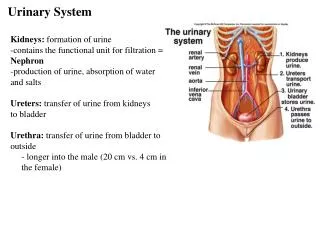
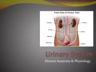
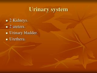
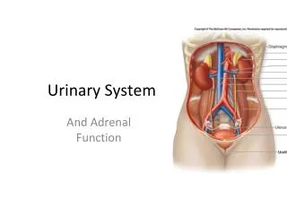
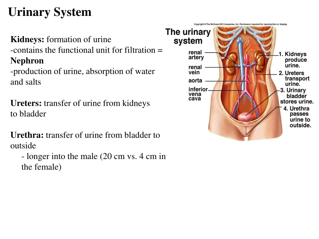
Urinary System. Kidneys: formation of urine -contains the functional unit for filtration = Nephron -production of urine, absorption of water and salts Ureters: transfer of urine from kidneys to bladder Urethra: transfer of urine from bladder to outside

E N D
Urinary System Kidneys: formation of urine -contains the functional unit for filtration = Nephron -production of urine, absorption of water and salts Ureters: transfer of urine from kidneys to bladder Urethra: transfer of urine from bladder to outside - longer into the male (20 cm vs. 4 cm in the female)
Kidneys • 10-12 cm • retroperitoneal – behind the peritoneum • not part of the abdominal cavity • surrounded by three layers of tissue: • 1. deepest layer = renal capsule – transparent sheet of dense irregular connective tissue • continuous with the outer coat of the ureter • 2. middle layer = adipose capsule • amass of fatty tissue surrounding the renal capsule • 3. outer layer = renal fascia • thin layer of dense irregular connective tissue that anchors the kidney to the abdominal wall
Kidneys • divided internally into an outer cortex and an inner medulla • medulla consists of 8 to 18 cone-shaped regions called renal pyramids • the wider base faces towards the cortex, the narrow region (renal papilla) projects down into a cup-like structure called a minor calyx • renal cortex is divided into an outer cortical zone and a deeper juxtamedullary zone • the cortex also extends down in between the pyramids to form the renal columns • renal lobe = renal pyramid + the overlying renal cortex + ½ the adjacent renal column
Blood supply • supplied by a renal artery and drained by a renal vein(s) • kidney receives 20-25% of the resting cardiac output through the renal arteries (1200mL per minute) • renal artery divides into segmental arteries – supply segments of the kidney • the segmental arteries give off branches that pass through the renal columns – interlobar arteries • at the base on the renal pyramids – between the medulla and cortex – they are called arcuate arteries
Blood supply • divisions from the arcuate are called the interlobular arteries (pass between the renal lobes) • the afferent arteriolesare derived from the interlobular arteries • afferent arteriole supplies one nephron and forms the glomerulus(capillary network) • drainage of the glomerulus is via the efferent arteriole
Blood supply • drainage of the glomerulus is via the efferent arteriole • efferent arteriole forms the peritubular capillary networkwhich surround the upper portions of the nephron • an extension of this network covers the lower portion of the nephron – vasa recta • the peritubular capillaries form the interlobular veins – arcuate veins – interlobar veins – renal vein
The Nephron -functional unit of the kidney -about one million nephrons between the two kidneys -filter 180 L fluid per day!!!! -each nephron is comprised of a renal corpuscle + renal tubules -renal corpuscle: filtering capsule surrounding the glomerulus -renal tubules: for reabsorption of water and ions leading to final urine volume and composition
The Nephron interlobular interlobular
Cortical Nephron • 80-85% of nephrons are cortical nephrons • Renal corpuscles are in outer cortex and majority of loops of Henle lie mainly in cortex
Juxtamedullary Nephron • 15-20% of nephrons are juxtamedullary nephrons • Renal corpuscles close to medulla and long loops of Henle extend into deepest medulla enabling excretion of dilute or concentrated urine
the blood capillaries covering the loop of Henle are called the vasa recta • have an important role in creating dilute or concentrated urine
Urinary System Function 1. Excretion of Metabolic Wastes: nitrogenous wastes -Urea: by-product of amino acid metabolism -produced when ammonia + carbon dioxide -Creatinine: produced by breakdown of creatine phosphate (high energy molecule reserve of muscles) -Uric acid: by-product of nucleotide breakdown -insoluble and ppts in the blood, concentrates in joints 2. Water-Salt balance of blood: reabsorption of sodium leads to the osmotic reabsorption of water from many parts of the nephron -”where sodium goes water follows” 3. Acid-Base balance of blood: reabsorption of bicarbonate ions from urine in the nephron decreases levels in blood (decreases carbonic acid levels) -movement of hydrogen ions from blood into the nephron, combines with ammonia to form ammonium (NH4+)
4. Secretion of hormones: release of renin by kidneys which leads to release of aldosterone by adrenal glands (reabsorption of salts by kidneys) -release of erythropoietin by kidneys (stimulates RBC production) -activation of vitamin D produced by the skin
Water balance • extracellular fluids: blood plasma & interstitial fluid • contain sodium, chloride, bicarbonate ions, potassium, calcium, magnesium, phosphate and sulfate ions • intracellular fluid: cytosol • higher potassium, phosphate, magnesium vs. ECF • lower sodium, chloride and bicarb ions vs. ECF • water is divided between our ECF and our ICF • of the 40 liters of water in the average male - 37% is ECF and 63%is ICF • blood pressure and osmotic pressure work together to control the movement of water in between the blood plasma and the interstitial flui • e.g. blood pressure causes the movement of fluid out of the arteriolar ends of capillaries into the interstitial fluid BUT osmotic pressure causes fluids to leave the interstitial fluid and enter the venular end of capillaries (i.e. BULK FLOW) • the excretory system’s ability to control the composition of blood plasma affects the composition of interstitial fluids which affects our composition of intracellular fluids
Renal physiology • comprised of filtration at the capsule (1) • reabsorption through the tubules (2) • direct secretion by the cells lining these tubules (3)
The Glomerulus • capillary tangle derived from afferent arterioles (into) and lead into efferent arterioles (out)
The Glomerulus • surrounded by a glomerular capsule (Bowman’s capsule) – made up of a: • parietal layer • visceral layer = single layer of epithelial cells known aspodocytes • space between the visceral and parietal layers = glomerular capsule • glomerular capsule: site of initial filtration and the first step in the formation of urine • the podocytes wrap around the endothelial cells of the glomerular capillaries and forms a filtration membrane together with the endothelial cell wall
The Glomerulus • the endothelial lining of the glomerulus itself contains “holes” for the passage of materials • the filtration membrane is really these holes + the “slits” between the podocytes
Glomerular Filtration • depends on three main pressures • 1. glomerular blood pressure (GBP)– BP in the glomerular capillaries • 55 mmHg (outward) • promotes filtration by forcing water and solutes through the filtration membrane at the glomerulus • 2. caspsular hydrostatic pressure (CHP)– hydrostatic pressure exerted back against the filtration membrane by fluid already in the Bowman’s capsule • opposes filtration from the blood • 15 mm Hg (inward) • 3. blood colloid osmotic pressure (BCOP)– inward force due to the presence of plasma proteins in the blood • creates an osmotic pressure in the blood plasma • opposes filtration from the blood • 30 mmHg (inward)
Glomerular Filtration • net filtration pressure (NFP) = GBP – (CHP + BCOP) • 10 mm Hg – net outward • filtration of plasma proteins into the urine/filtrate can at the glomerulus can increase NFP • damage to the glomerular capillaries can increase their permeability – loss of the larger plasma proteins into the urinary filtrate – not reabsorbed through the rest of the nephron due to large size • this increases the osmotic pressure in the urinary filtrate which draws larger amounts of water out of the blood and into the urine filtrate • but the BCOP decreases in the blood plasma because we are losing these plasma proteins in the urine – increases NFP
Glomerular filtration rate • glomerular filtration rate (GFR) = amount of filtrate formed per minute • (125 mL/min) • affected dramatically by NFP • adjusted by regulating: • 1) blood flow into and out of the glomerulus • 2) the glomerular capillary surface area available for reabsorption • three mechanisms control GFR
GFR Control Mechanisms • 1. renal autoregulation • designed to keep renal blow flow and GFR reasonably constant • without it – even a small change in renal blood pressure would cause a significant change in GFR • two mechanisms: myogenic mechanism and tubuloglomerular feedback
GFR Control Mechanisms • 1. renal autoregulation - myogenic mechanism • increased blood volume/pressure can increase GFR • the stretching of the afferent arterioles triggers the contraction of the smooth muscle lining these arterioles = vasoconstriction • narrowing of the arterioles lumen decreases blood flow and reducing the GFR • smooth muscle lining the efferent arteriole relaxes vasodilation
GFR Control Mechanisms • 1. renal autoregulation - tubuloglomerular mechanism • feedback provided to the glomerulus from the renal tubules • with elevated GFR there is an increase in the fluid flowing through the PCT, LH and DCT • increased flow rate results - less time to reabsorb materials • macula densa cells surrounding the afferent arteriole induce vasoconstriction in the afferent arterioles • if GFR drops below normal – these cells stimulate the release of nitric oxide from the juxtaglomerular cells vasodilation which increases blood flow and GFR
GFR Control Mechanisms • 2. neural regulation – sympathetic ANS fibers release norepinephrine which causes vasoconstriction of the smooth muscle in the afferent arteriole • at rest norepinephrine release is low • with increased sympathetic activity – decreased GFR • 3. hormonal regulation • release of angiotensin II reduces GFR by inducing vasoconstriction • release of atrial natriureic peptide (ANP – from the atrium) increases GFR by increasing the surface area of the glomerulus • stretching of the atria as blood blow increases stretches the cells and induces the release of ANP • ANP relaxes the mesangial cells and increases the SA of the glomerular capillaries – increased GFR
Renal Physiology • Tubular reabsorption • tubule cells reabsorb about 99% of the filtered water and many of the solutes • principal materials reabsorbed – glucose, amino acids, urea, Na+, K+, Ca+, Cl-, HCO3- and HPO4- • return to the blood through reabsorption into the peritubular capillary network and vasa recta • reabsorption= return to the blood • absorption = entrance of new materials into the blood (e.g. via digestive absorption)
Reabsorption Routes basal face apical face • reabsorption routes – one of two routes before re-entering the blood • Paracellular reabsorption • in some parts oftubule - 50% of reabsorbed materialmoves between cells bydiffusion • apical face of cells linked by “leaky” tight junctions • Transcellular reabsorption • material moves throughboth the apical and basalmembranes of the tubulecell by active transport
Renal Physiology • Tubular secretion • tubular cells also secrete other materials – wastes, drugs, excess ions into the urine • this also removes these materials from the blood
Reabsorption in the 1st half of PCT • Intracellular sodium levels are kept low due to primary active transport - Na+/K+ pump • creates a sodium gradient across the apical and the basal faces • Na+ symporters help reabsorb materials from the tubular filtrate as sodium ions diffuse into the cell • glucose, amino acids, lactic acid, water-soluble vitamins and other nutrients are completely reabsorbed in the first half of the proximal convoluted tubule
Reabsorption of Bicarbonate, Na+ & H+ Ions • Na+ antiporters reabsorb Na+ from the filtrate and secrete H+ into the filtrate • PCT cells produce the H+ from the dissociation of H2CO3
Reabsorption of Bicarbonate, Na+ & H+ Ions • For every H+ secreted into the tubular fluid, one filtered bicarbonate eventually returns to the blood via diffusion • PCT cells release bicarbonate ion to the peritubular capillaries • important buffering system of blood
Passive Reabsorption in the 2nd Half of PCT • Electrochemical gradients produced by symporters & antiporters causes passive reabsorption of other solutes • Cl-, K+, Ca+2, Mg+2 and urea passively diffuse into the interstitial space and then into the peritubular capillaries • promotes osmosis in PCT - especially permeable due to aquaporin channels
Secretion of NH4+ in PCT • Ammonia (NH3) is a poisonous waste product of protein deamination by the liver • most is converted to urea which is less toxic • both ammonia & urea are filtered at the glomerulus • NH3 is also created by the cells of the PCT • PCT cells deaminate glutamine - generates both NH3 and a new bicarbonate ion. • the ammonia is combined with H+ to create ammonium • secreted into the filtrate by a Na+ antiporter • Bicarbonate is moved with Na+ by a symporter into the interstitial space • diffuses into the bloodstream
Reabsorption in the Loop of Henle • Tubular fluid • PCT reabsorbed 65% of the filtered water so chemical composition of tubular fluid in the loop of Henle is quite different from plasma • since many nutrients were reabsorbed as well, osmolarity of tubular fluid is close to that of blood • Sets the stage for independent regulation of both volume & osmolarity of body fluids
Symporters in the Loop of Henle • Thick, ascending limb of loop of Henle has Na+ K- Cl- symporters that reabsorb these ions in response to a sodium gradient • K+ leaks back through K+ channels into the tubular filtrate • Cl- diffuses into the interstitial fluid and blood • results in a negatively charged fluid • Cations passively move into the vasa recta
Countercurrent Mechanism of the Loop of Henle • Descending limb is very permeable to water • higher osmolarity of interstitial fluid outside the descending limb causes water to mover out of the tubule by osmosis • at hairpin turn, osmolarity can reach 1200 mOsm/liter • Ascending limb is impermeable to water • but symporters remove Na+ and Cl- & establish as osmotic gradient for the reabsorption of water from the descending limb
Countercurrent Mechanism • Na+ is pumped into the • interstitial space between the limbs • Causes a temporary increase • in osmotic pressure within the interstitial • space • Increase in OP causes water • to be “sucked out” of filtrate moving down the descending limb • this makes the filtrate in the ascending limb “saltier” – more Na+ to be pumped into the interstitial space • can also cause water to move out of DCT and collecting duct (but ADH must be produced for this to happen)
DCT and Collecting Duct • two types of cells found in the DCT and CD • principal cells – receptors for ADH and aldosterone • intercalated cells – play a role in the homeostasis of blood pH • DCT and collecting duct are impermeable to water !!!! • the DCT and CD become permeable upon action of hormones
Reabsorption in the DCT • Removal of Na+ and Cl- continues in the DCT by means of Na+ pumps and Na+ Cl- symporters • Na+ and Cl- then reabsorbed into peritubular capillaries • NO reabsorption of water unless hormones are produced • DCT is major site where parathyroid hormone stimulates reabsorption of Ca+2 • DCT is not very permeable to water so it is reabsorbed with little accompanying water
Actions of the Principal Cells • Na+ pumps create a sodium gradient • Na+ enters principal cellsthrough leakage channels • in exchange cells secrete variable amounts of K+ into the filtrate & interstitial fluid • to adjust for dietary changes in K+ intake • down concentration gradient due to Na+/K+ pump Aldosterone • Aldosterone increases this Na+ reabsorption & K+ secretion by principal cells by stimulating the synthesis of new pumps and channels.
Actions of the Principal Cells • ADH increases the expression of aquaporin-2 channels in the cell membrane – increased reabsorption of water when dehydrated Aldosterone H20 (ADH)
Secretion of H+ and Absorption of Bicarbonate by Intercalated Cells • combination of CO2 and H20 creates H2CO3 H+ and HCO3- • proton pumps (H+ATPases) secrete H+ into tubular fluid • can secrete against a concentration gradient so urine can be 1000 times more acidic than blood • Cl-/HCO3- antiporters move bicarbonate ions into the interstitial space and then the blood • so intercalated cells help regulate pH of body fluids
Buffering of Urine by Intercalated Cells • Urine is buffered by HPO4 2- and ammonia - both of which combine irreversibly with H+ and are excreted • H+ in the filtrate a pumped there by H+ ATPase pups
Reabsorption & Secretion in the Collecting Duct • By end of DCT, 95% of solutes & water have been reabsorbed and returned to the bloodstream • Cells in the collecting duct make the final adjustments • principal cells reabsorb Na+ and secrete K+ • intercalated cells reabsorb K+ & bicarbonate ions and secrete H+
Production of Dilute or Concentrated Urine • Homeostasis of body fluids despite variable fluid intake • Kidneys regulate water loss in urine • ADH controls whether dilute or concentrated urine is formed • if lacking, urine contains high ratio of water to solutes
Summary • H2O Reabsorption • PCT---65% • loop---15% • DCT----10-15% • collecting duct--- 5-10% with ADH
Renin-Angiotensin-Aldosterone • when blood volume and BP drop – the walls of the afferent arterioles are stretched less • juxtaglomerular cells secrete renin into the blood (also stimulated by sympathetic stimulation) • in the blood renin cleaves angiotensinogen (made by hepatocytes) to form the active enzyme angiotensin I • the enzyme ACE (expressed by endothelial cells) – cleaves this even more to form angiotensin II • the lung is a great source of ACE due to its density of capillaries
Renin-Angiotensin-Aldosterone • angiotensin II actions: • 1. decreases GFR by causing vasoconstriction of afferent arterioles • 2. enhances reabsorption of Na+, Cl+ and water in the PCT by stimulating the Na/H antiporter • 3. stimulates the release of aldosterone by the adrenal cortex • aldosterone stimulates the principal cells of the DCT & collecting ducts to reabsorb more Na+ and Cl- and secrete more K+ into the blood • osmotic consequence of this causes an increased reabsorption of water throughout the body
ADH ADHreceptor • an increase in osmolarity in the blood plasma triggers the release of ADH, which helps the kidney to conserve water • ADH – released by the posterior pituitary • regulates water reabsorption by increasing the permeability of the principal cells to water • increases synthesis of aquaporin channels • when the OP of the blood plasma increases due to dehydration – osmoreceptors in the hypothalamus detect this drop and stimulate the release of ADH • ADH causes increased reabsorption of water from the urine through the aquaporin channels LUMEN COLLECTINGDUCT CELL ADH cAMP Second-messengersignaling molecule Storagevesicle Exocytosis Aquaporinwater channel H2O H2O