Download
1 / 69
1.07k likes | 2.13k Views
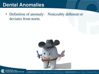
Dental Anomalies in Radiology. Developmental VS. acquired . Developmental Abnormalities. Supernumerary Teeth (hyperdontia, supplemental teeth). 1~4% , familial tendency Mesiodens, paramolar Distodens, distomolar teeth Peridens Single : premaxilla, maxillary molar

E N D
Dental Anomalies in Radiology Developmental VS. acquired
Supernumerary Teeth (hyperdontia, supplemental teeth) • 1~4% , familial tendency • Mesiodens, paramolar • Distodens, distomolar teeth • Peridens • Single : premaxilla, maxillary molar • Multiple : premolar area, mandibular • M : F = 2 : 1 • Impaction or delay eruption of normal teeth; dentigerous cyst Syndrome: • Cleidocranial dysplasia, Gardner’s syn.
Missing Teeth • 3~10%, excluding 3rd molars • Hypodontia • Oligodontia • Anodontia • 8 > 5 > 2 > 1 • Ectodermal dysplasia ; orofaciodigital syndrome
Q: 請就以上同一名患者的根尖X光片,說明有何異常。
SIZE OF TEETH • True generalized type and relative type Macrodontia • Hemangioma, hemihypertrophy of the face, pituitary giantism Microdontia • pituitary dwarfism • supernumerary teeth, 3rd molars, lateral incisors
Macrodontia Microdontia
ERUPTION OF TEETH Transposition • Two teeth exchanged positions • 3 & 4 ; 3 & 2, 657
Gemination (twinning) -Division of a single tooth bud • primary dentition , esp. incisor region • complete twinning increase tooth number • pulp chamber is single & enlarged, maybe partial divided
Fusion (synodontia) • bifid crown or two recognizable teeth, reduced number of teeth • more common in the primary dentition, esp. anterior region -Adjacent tooth germs combined with dentin or enamel
Concresence - Roots of two or more teeth united by cementum • space restriction during develop., local trauma, excessive occlusal force or local infection after development • maxillary molars; 3rd molar & a supernumerary tooth
Fusion / Gemination • A tooth with two separated root canals and with one or two roots…Fusion • An enlarged tooth with a bifid crown containing an enlarged or possibly partially divided pulp chamber…Gemination
Taurodontism -Longitudinal enlarged pulp chamber, increased distance between CEJ to the bifurcation • normal crown size & tooth length, shortened roots • not recognizable clinically • most in molars • Trisomy 21
Dilaceration • A sharp bend or curve in the crown or root • maxillary premolars
Dens in Dente(dens invaginatus) - Infolding of the outer enamel surface into the interior • at the anatomically defined pit • caries→pulpal disease
coronal type: enamel organ infolding into the dental papilla; 2>1>4,5>3 • radicular type: invagination of Hertwig’s epithelial root sheath, lined with cementum; • 4, 7
radicular type Dilated odontome
Dens Evaginatus - Outfolding of enamel organ • a tubercle on occlusal surface, with enamel surface & dentin core, pulp horn often extends into the evagination • premolar or molar • pulp infection due to fracture
Lingual pits Dens Evaginatus
Amelogenesis Imperfecta -Disturbance in enamel development • Normal dentin & root • autosomal dominant or recessive , X-linked • Four general types
1.Hypoplastic type • Thin enamel with pitted, rough or smooth & glossy surface; yellowish to brown • undersized, squared crown, lack of contact • flat occlusal surface & low cusps, attrition
2.Hypomaturation • normal thickness of enamel, but mottled surface; cloudy white, yellow or brown, opaque in color • softer than normal • same density as dentin
3.Hypocalcified type • normal thickness of enamel, density less than dentin • normal size & shape when erupt, abrade or fracture away rapidly • permeability increase, darkened & stained 4.Hypomaturation-hypocalcified with taurodontism
Dentinogenesis Imperfecta (hereditary opalescent dentin) • autosomal dominant hereditary Type I : D.I. + osteogenesis imperfecta Type II : D.I., no skeletal defects • enamel fractures, attrition severely • dark brown to black
Dentinogenesis Imperfecta Osteogenesis imperfecta
Radiographic Features of D.I. • bulbous crown, normal size, constriction of the cervical area • short & slender roots • occlusal attrition • partial or complete obliteration of the pulp chambers, root canals absent or threadlike
Dentinogenesis Imperfecta
Dentin Dysplasia -autosomal dominant disturbance • rare (1:100,000) Type I (radicular) • normal color & shaped in both dentition • malaligned arch, drifting and exfoliate with little or no trauma • short or abnormal root shaped, pulp chamber & root canals completely fill in before eruption • 20 % of teeth with type I disease have apical radiolucencies
TypeII (coronal) • primary dentition appears as D.I., but permanent dentition is normal • obliterated of the pulp chamber & reduced root canals after eruption • roots are normal in shape & proportion
Regional Odontodysplasia (odontogenesis imperfecta) - hypoplastic & hypocalcified of both dentin & enamel • only a few adjacent teeth in a quadrant affected either primary or permanent teeth • central incisors > lateral incisors >canines (maxillary) • delayed eruption • ghostlike appearance in image • large pulp chamber & wide root canals, roots are short & poorly outlined • thin enamel , less dense as usual
Enamel Pearl (enameloma, enamel drop, enamel nodule) - small globule of enamel on the roots furcation area of molars • prevalence : 3 % • mesial or distal aspect in Max. molar and buccal or lingual in Mand. molars
Talon Cusp - Anomalous hyperplasia of the cingulum of a Max. or Mand. incisor →a supernumerary cusp • T shaped in incisal view • Differential diagnosed with supernumerary tooth
Turner’s Hypoplasia (Turner’s tooth) -a local hypoplastic or hypomineralized defect in crown of a permanent tooth • extension of a periapical infection or mechanical trauma from deciduous predecessor • most common in lower premolars
Congenital Syphilis • 30 % p’t develop dental hypoplasia • Hutchinson’s incisors & mulberry molars • not all p’t with Hutchinson’s teeth or mulberry molars will have congenital syphilis
Congenital syphilis Hutchinson’s incisors & mulberry molars