Download
1 / 1
10 likes | 150 Views
Characterization of Macroporous Monolithic Poly (2- Hydroxyl ethyl Methacrylate) Cryogels W. Akande*, L. Mikhalovska, I. Savina, S. James, S. Mikhalovsky School of Pharmacy and Biomolecular Sciences, University of Brighton, Brighton, UK Corresponding author: wa20@brighton.ac.uk.

E N D
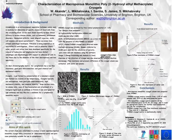
Characterization of Macroporous Monolithic Poly (2- Hydroxyl ethyl Methacrylate) Cryogels W. Akande*, L. Mikhalovska, I. Savina, S. James, S. Mikhalovsky School of Pharmacy and Biomolecular Sciences, University of Brighton, Brighton, UK Corresponding author: wa20@brighton.ac.uk Introduction & Background Methods Results Cytapheresis is an extracorporeal separation technique widely used in medicine for elimination of specific classes of blood cells from the circulating blood. It has been shown recently to have clinical efficacy in various disease states, such as leukaemia, autoimmune disorders, rheumatoid arthritis, renal allograft rejection, sickle – cell anaemia and severe parasitemia[1]. These successes have utilised centrifugal techniques. Filtration is being explored as an alternative to centrifugation. Filters such as polyester fibers, nylon, acrylic and cotton have been developed specifically for the removal of leukocytes and lymphocytes but have distinct advantages and disadvantages with respect to adhesion of cells to the fibers due to the diameter of the fiber (microporous) and less specificity. An ideal chromatography matrix for cytapheresis has a low flow résistance; good pore interconnection; and good chemical and mechanical stability. Cryogels, a gel formed by polymerisation of a monomers around pre-formed ice crystals at low temperature2. Cryogels typically are macroporous, have good pore interconnectivity, high mechanical stability, and chemical stability, large surface area-to-volume ratio, ease of functionalisation and attachment of a biological ligand such as antibody or Protein A (an avid ligand for IgG antibody) and may thus be a candidate for a cytapheresis monolithic matrix PHEMA cryogel was produced by free radical polymerisation at -12oc for 18hours from monomers i)2-hydroxylethyl methacrylate (HEMA) and ii)allyl glycidyl ether (AGE) iii) N, N1methylenebisacrylamide (MBAA) as a cross linker. iv)At the presence of APS/TEMED as initiator v)FITC stained PHEMA cryogel were observed under Confocal microscope (CLSM). Images Collected by CLSM were used for the calculation of porosity, pore sizes and wall thickness using the software Image J (Image Processing and Analysis in Java). Overview of the structure of cryogel was also observed by scanning electron microscopy. Flow resistance and pressure difference of the cryogel column was estimated with water and plasma . cryogel in 5cm x 1cm glass columns Figure 5:Flow resistance of PHEMA cryogel Results Figure 6: flow rate /∆pressure of water and plasma Conclusions • Macroporous monolithic cryogel column was prepared from PHEMA • PHEMA cryogel matrix consists from ~13% of solid polymer and ~87% of the interconnected pores • The monolithic cryogel matrix consists of the interconnected pores of 41-81 mm range, and it could be appropriate matrix for further functionalisation and use for cytapheresis • PHEMA cryogel has a low flow resistance evaluated at flow rate of 1-20ml/min • PHEMA cryogel matrix is mechanically stable Fig 1. SEM of PHEMA cryogel Figure 2. Confocal Microscopic images of PHEMA cryogel Whole blood containing cells A,B and C Porosity, pore size (Fig. 3) and wall thickness of the PHEMA cryogel matrix were calculated from the CLSM images. Cell (B) specific cryogel matrix Cell A Whole blood containing cells A and C References Onodera H, Ninomiya K, Yoshida M, et al. (2003) Therap Apher 7(3): 329. Plieva FM, et al. J Separation Sci (2004) 27: 828 Cell B Cell C Diagrammatic expression of a cell specific cytapheresis column Acknowledgements This work was funded by the FP7 MONACO project Aims of this work The current study was undertaken to produce a novel supermacroporous monolithic cryogel with a structure of interconnected pores and pore sizes potentially suitable for use in cytapheresis. Figure 4 :Graph of flow rate of water versus ∆Pressure in 8% PHEMA cryogel Figure 3: Pore size distribution of PHEMA cryogel matrix