Download
1 / 7
70 likes | 373 Views
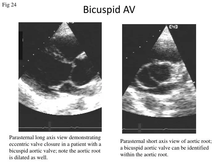
Fig 24. Bicuspid AV. Parasternal long axis view demonstrating eccentric valve closure in a patient with a bicuspid aortic valve; note the aortic root is dilated as well. Parasternal short axis view of aortic root; a bicuspid aortic valve can be identified within the aortic root. .

E N D
Fig 24 Bicuspid AV Parasternal long axis view demonstrating eccentric valve closure in a patient with a bicuspid aortic valve; note the aortic root is dilated as well. Parasternal short axis view of aortic root; a bicuspid aortic valve can be identified within the aortic root.
Fig 25 Bicuspid aortic valve causing aortic stenosis Parasternal long axis view in a patient with a bicuspid aortic valve, demonstrating thickened valve leaflets. Doppler colour flow demonstrating the proximal acceleration and distal flow disturbance.
Fig 26 Bicuspid aortic valve with AR RV Ao LV Ao LV LA LA Parasternal long axis view in a patient with a bicuspid aortic valve, demonstrating thin but doming aortic valve leaflets. Doppler colour flow demonstrating the aortic valve regurgitation during diastole.
Fig 27 Congenital AS RV Ao Ao LV Parasternal longaxis and short axis view showing a congenital aortic stenosis causing by a possible unileaflet aortic valve. The valve leaflet itself looks dysplastic.
Fig 28 Sub AS b a LV RV LV vsd RV Ao LA c a:Apical five chamber view demonstrating a sub-aortic ridge. b:Colour flow Doppler showing turbulent flow start from sub-aortic area and a shunt through a VSD. c: Continuous Doppler trace from the LVOT showing a peak velocity of 4.2 m/s
Fig 29 Sub aortic stenosis Ao LV 3D imagines demonstrating a sub-aortic fibrio-muscular ring
Fig 30 Small Aortic Root Colour Doppler demonstrate mild aortic regurgitation Parasternal long-axis view of a fibromuscular subaortic tunnel. The aortic root itself is also small