Download
1 / 43
440 likes | 594 Views
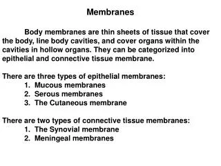
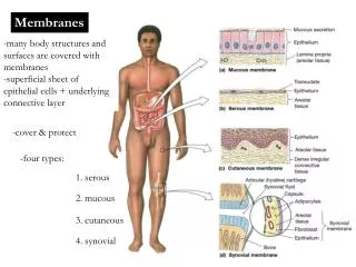
Cellular membranes. 2 / 16. Overview of the body. 3 / 16. The cell. 4 / 16. Biological membranes. the surface of the cells and the organelles are covered with membranes – compartmentalization

E N D
2/16 Overview of the body
3/16 The cell
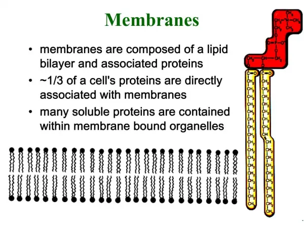
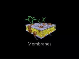
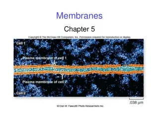
4/16 Biological membranes • the surface of the cells and the organelles are covered with membranes – compartmentalization • Karl Wilhelm von Nägeli middle of the XIX. century – there is a barrier against movement of pigments on the surface of cells – swelling and shrinking - plasma membrane • direct proof only with EM • Singer and Nicholson (1972): fluid mosaic hypothesis • 6-8 nm lipid bilayer + proteins • mosaic, because proteins tend to group • fluid, because they can easily move laterally • lipid/protein ratio depends on function: myelin and mitochondrion • 106 lipid molecules/μ2 Eckert: Animal Physiology, W.H.Freeman and Co., N.Y.,2000, Fig. 4-2.
5/16 Lipid components I. • phospholipids • usually more then half of total lipid content • phosphoglycerides • phosphatidylcholine (lecithin) • phosphatidylserine • phosphatidylethanolamine • other, e.g. phosphatidylinositol (PI, PIP, PIP2) • role of the cis-, and trans conformation • sphingomyelins • serine + fatty acid = sphingosine (condensation of COOH groups) • sphingosine + fatty acid = ceramide (on the amino group of serine) • ceramide + phosphate + choline = sphingomyelin (on the OH group of serine) Alberts et al.: Molecular biology of the cell, Garland Inc., N.Y., London 1989, Fig. 12-21. Eckert: Animal Physiology, W.H.Freeman and Co., N.Y.,2000, Fig. 4-3. Alberts et al.: Molecular biology of the cell, Garland Inc., N.Y., London 1989, Fig. 6-9. Alberts et al.: Molecular biology of the cell, Garland Inc., N.Y., London 1989, Fig. 6-9.
glycolipids on the outer surface only cell to cell recognition, antigens (e.g. blood types) plants and bacteria: based on glycerol animals: based on ceramide neutral: e.g. galactocerebroside (serine OH in ceramide binds galactose builds up 40% of myelin outer membrane gangliosides (serine OH in ceramide binds oligosaccharide containing one or more charged sialic acid (N-acetylneuraminic acid - NANA) 5-10% f total lipids in nerve cells steroids cholesterol mainly more than 18% decreases fluidity, inhibits crystallization 6/16 Lipid components II. Alberts et al.: Molecular biology of the cell, Garland Inc., N.Y., London 1989, Fig. 6-13. Darnell et al., Scientific American Books, N.Y., 1986, Fig. 3-79 Darnell et al., Scientific American Books, N.Y., 1986, Fig. 14-32 Eckert: Animal Physiology, W.H.Freeman and Co., N.Y.,2000, Fig. 4-7. Eckert: Animal Physiology, W.H.Freeman and Co., N.Y.,2000, Fig. 4-4. Alberts et al.: Molecular biology of the cell, Garland Inc., N.Y., London 1989, Fig. 6-11.
7/16 Protein components • integral or intrinsic proteins: embedded in the membrane, reaching from one side to the other • transmembrane part usually forms -helix, with hydrophobic side chains on the outside • transmembrane parts can be predicted by the sequence of amino acids (hydrophobicity) • often multiple transmembrane parts: e.g. 7TM receptors • helices are connected by loops • functions: ion channel, receptor, enzyme, transporter, etc. • peripheral or extrinsic proteins: associated with the membrane on one side only • they can be enzymes, proteins serving signalization (G-proteins), etc.
8/16 Membrane as a barrier • the membrane prevents free exchange of materials - compartmentalization • classification by substances: • hydrophobic (non-polar) substances - diffusion • hydrophilic (polar) substances • uncharged: • small molecular weight – diffusion • higher molecular weight – by carrier molecules • ions – through ion channels • classification by use of energy: • passive: along the gradient – energy is not needed (diffusion, facilitated diffusion, channel) • active: against the gradient – direct or indirect use of energy – transport molecules • special: endocytosis, exocytosis Eckert: Animal Physiology, W.H.Freeman and Co., N.Y.,2000, Fig. 4-18.
9/16 Diffusion I. • difference between convection (bulk flow) and diffusion • water molecules travel 2000 km in one hour, but in random directions • glucose only (?) 700 km/h • time changes by the square of time • example: glucose in capillary: • 10 - 90% - 3,5 s 10 cm - 90% - 11 years • size limit for cells (30-50 ), plasma flow, axonal transport systems • Fick’s first law: J = -D*A*dc/dx • flow and concentration is considered from a given point into x-direction
10/16 Diffusion II. • for spherical molecules (Stokes-Einstein relation):D = kT / (6 r) • diffusion through a lipid layer depends on concentration at the edges of the lipid layer • it depends on the partition coefficient as concentration in the water phase is constant • thus the gradient is given by:K(co - ci) / x consequently J = - DmKA (co - ci) / x • partition and diffusion coefficients as well as membrane width are constant for any given substance – permeability coefficient is defined J = - PA (co - ci) • related parameter: conductance
11/16 Osmosis I. • in fact it is the diffusion of water • penetrates easily, water compartments are in equilibrium • Abbé Jean Antoine Nollet (1748) described it first experimenting with a bladder • to reach equilibrium, hydrostatic pressure is needed on the side of the solution – osmotic pressure • osmos (Greek) = to push • linear relationship with temperature (T) and osmolarity (particles per liter of solvent) • van’t Hoff: molecules in solution behave thermodynamically like gas molecules • volume of 1 mol gas at room temperature is 24 liters • osmotic pressure of a solution of 1 osmole is 24 atm at room temperature
12/16 Osmosis II. • osmotic pressure depends on the number of particles: = i * m * RT • it is usually calculated from molarity using a correction factor taken from precalculated tables • it is measured by changes in freezing and boiling points • hyposmotic, hyperosmotic, isosmotic • hypotonic, hypertonic, isotonic • similar but not equivalent notions! • first is calculated, second is observed as the effect on living cells, e.g. glycerol and NaCl • isosmotic NaCl solution: saline (0,9%), physiological solution
13/16 Ion channels • built up by intrinsic (integral) proteins • -helices, connected by loops • ions (Na+, K+, Ca++, Cl-, etc.) can only pass through channels or by transport molecules • analysis using patch clamp method • selectivity for ions – size, charge, dehydration energy (K+ > Na+) • large families: grouped by ion specificity and opening mode • leakage, voltage-, ligand-dependent, mechanosensitive • voltage-dependent: best known: 4 motifs, 6 helices each - Na+, Ca++ 1 protein molecule, K+ 4 molecules, with 1-1 motif ; three states • ligand-dependent: 5 motifs (pentamer) in general, 5 molecules, each with 4 helices Alberts et al.: Molecular biology of the cell, Garland Inc., N.Y., London 1989, Fig. 6-58. Alberts et al.: Molecular biology of the cell, Garland Inc., N.Y., London 1989, Fig. 6-64. Eckert: Animal Physiology, W.H.Freeman and Co., N.Y.,2000, Fig. 4-30. Alberts et al.: Molecular biology of the cell, Garland Inc., N.Y., London 1989, Fig. 6-60, 6-61. Eckert: Animal Physiology, W.H.Freeman and Co., N.Y.,2000, Fig. 5-28.
14/16 Transport by carriers I. • conformation change upon binding of the transported molecule • do not travel between the two sides of the membranes • grouped by the use of energy: • facilitated diffusion • active transport • grouped by the number of carried substances • uniporter – 1 substance • symporter - 2 substances in the same direction • antiporter - 2 substances in opposite directions • characteristics: • saturation • selectivity • competition Eckert: Animal Physiology, W.H.Freeman and Co., N.Y.,2000, Fig. 4-23.
15/16 Transport by carriers II. • facilitated diffusion • along the gradient • no use of energy • large, polar molecules, e.g. glucose • active transport • direct use of energy, hydrolysis of ATP • in the case of ions, it is called a pump • Na + /K + pump, in neuronal and muscle cells - antiporter - exact mechanism is not known • H+ - mitochondrion - ATP synthesis by the passage of 3 H+ • indirect use of energy, usually on the expense of the Na+ gradient • e.g. uptake of glucose and amino acids in the kidney and gut - gradient is small • water uptake in the kidney Eckert: Animal Physiology, W.H.Freeman and Co., N.Y.,2000, Fig. 4-24. Eckert: Animal Physiology, W.H.Freeman and Co., N.Y.,2000, Fig. 4-40.
16/16 Endocytosis and exocytosis • transport of macromolecules • endocytosis – uptake of substances • mechanism: vesicle budding off from the membrane • pinocytosis – “drinking” – small vesicles – constitutive, continuous in all cells – e.g. membrane recycling • phagocytosis – “eating” – larger vesicles stimulus-induced, in special cells • receptor-mediated endocytosis • “clathrin coated pits” - receptors accumulate • units with lysosome after budding off • entrance of proteins, hormones, viruses, toxins, etc. • exocytosis – release of substances • mechanism: fusion of vesicle with the membrane • signal-induced exocytosis – nerve and endocrine cells – role of Ca++ • constitutive exocytosis – going on continuously Alberts et al.: Molecular biology of the cell, Garland Inc., N.Y., London 1989, Fig. 6-65. Eckert: Animal Physiology, W.H.Freeman and Co., N.Y.,2000, Fig. 4-31.
Fluid mosaic membrane Eckert: Animal Physiology, W.H.Freeman and Co., N.Y.,2000, Fig. 4-2.
Types of phospholipids Alberts et al.: Molecular biology of the cell, Garland Inc., N.Y., London 1989, Fig. 6-9.
Inositol phosphates Alberts et al.: Molecular biology of the cell, Garland Inc., N.Y., London 1989, Fig. 12-21.
Phosphoglycerides Eckert: Animal Physiology, W.H.Freeman and Co., N.Y.,2000, Fig. 4-3.
Glycocalyx Darnell et al., Scientific American Books, N.Y., 1986, Fig. 14-32
AB0 blood types Darnell et al., Scientific American Books, N.Y., 1986, Fig. 3-79
Cerebrosides Alberts et al.: Molecular biology of the cell, Garland Inc., N.Y., London 1989, Fig. 6-11.
Gangliosides Alberts et al.: Molecular biology of the cell, Garland Inc., N.Y., London 1989, Fig. 6-13.
Structure of cholesterol Eckert: Animal Physiology, W.H.Freeman and Co., N.Y.,2000, Fig. 4-4.
Cholesterol in the membrane Eckert: Animal Physiology, W.H.Freeman and Co., N.Y.,2000, Fig. 4-7.
Passing through the membrane Eckert: Animal Physiology, W.H.Freeman and Co., N.Y.,2000, Fig. 4-18.
Examination of ion channels Alberts et al.: Molecular biology of the cell, Garland Inc., N.Y., London 1989, Fig. 6-60, 6-61.
Selectivity of channels Eckert: Animal Physiology, W.H.Freeman and Co., N.Y.,2000, Fig. 4-30.
Voltage-dependent channels Eckert: Animal Physiology, W.H.Freeman and Co., N.Y.,2000, Fig. 5-28.
Activation - inactivation Alberts et al.: Molecular biology of the cell, Garland Inc., N.Y., London 1989, Fig. 6-58.
Nicotinic Ach receptor Alberts et al.: Molecular biology of the cell, Garland Inc., N.Y., London 1989, Fig. 6-64.
Transport types Eckert: Animal Physiology, W.H.Freeman and Co., N.Y.,2000, Fig. 4-23.
Facilitated diffusion Eckert: Animal Physiology, W.H.Freeman and Co., N.Y.,2000, Fig. 4-24.
Indirect active transport Eckert: Animal Physiology, W.H.Freeman and Co., N.Y.,2000, Fig. 4-40.
Receptor-mediated endocytosis Eckert: Animal Physiology, W.H.Freeman and Co., N.Y.,2000, Fig. 4-31.
Exocytosis in the synapse Alberts et al.: Molecular biology of the cell, Garland Inc., N.Y., London 1989, Fig. 6-65.