Download
1 / 38
380 likes | 388 Views
Basic concepts of Metabolism Metabolism and metabolic pathway Metabolic Map Catabolism Anabolism Regulation of Metabolism Signals from within the cell (Intracellular) Communication between cells. - Biosignaling: Signal transduction * Transduction by Intracellular receptors

E N D
Basic concepts of Metabolism • Metabolism and metabolic pathway • Metabolic Map • Catabolism • Anabolism • Regulation of Metabolism • Signals from within the cell (Intracellular) • Communication between cells. • - Biosignaling: Signal transduction • * Transduction by Intracellular receptors • * Transduction by Cell-surface receptors • a. Ligand-Gated Ion channels • b. Receptor enzyme • c. Receptors involving second messenger molecules • i. Adenylate cyclase system • ii. Phosphatidylinositol system • iii. Ca+ as second messenger • * Other messenger systems • a. cGMPs • b. Nitric oxide • References: • chapter 8 of Lippincots • chapter 13 of Lehningers
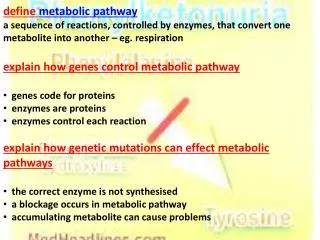
* Enzymes catalyze different reactions that don't occur in isolation but organized into multi step sequence called pathway. *The product of one reaction will be the substrate of the subsequent reaction * Different pathways intersect forming an integrated reactions collectively called Metabolism
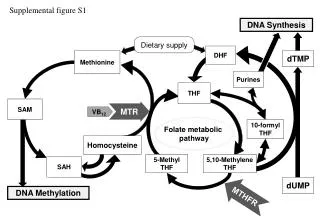
* Metabolic Map A picture containing the central pathways of energy metabolism. Each pathway is composed of multienzyme sequence and each enzyme has catalytic or regulatory features The Metabolic Map Shows the links between pathways, indicates the intermediates between different cycles (pathways) and shows the effect on different intermediates if one pathway is blocked
Catabolic and Anabolic Pathways Pathways can be classified as either catabolic (degradative) or anabolic (synthetic) Catabolic: degradation of complex molecules (polysaccharides, proteins) into simple molecules like CO2, NH3 and water. Catabolism is convergent process: a wide variety of molecules are transformed into a few common end products Anabolic reactions are the synthesis of complex molecules from simple precursor. Anabolic is divergent process in which few biosynthetic precursors form a wide variety of polymeric or complex products. Catabolism - catabolic reactions provide chemical energy in the form of the ATP from the degradation of the energy-rich fuel compound.The catabolism is essential for providing energy necessary for building up the complex compound
Energy generation occur in three steps Stage I Stage II Stage III Energy is librated from the transfer of electrons from NADH and FADH2 to O2 through the electron transport chain
Anabolism - Anabolic reactions combine small molecules as amino acids to form large complexes as proteins. These processes require energy which is provided by the break down of ATP to ADP - The biosynthetic pathway usually is different from degredative pathway of the same compound so the two processes respond to different regulatory - Anabolic reactions involve chemical reduction in which the reducing power is NADPH NADPH
Regulation of Metabolism • Pathways of the metabolism must be coordinated so that the catabolism and the anabolism must meet the needs of the cell. • The cell is not present in isolation it present in a tissue, in which all the cells communicate together with regulatory signals. • Regulatory signals including hormones, nervous system and availability of nutrients which affect the signals generated within the cell itself. • *Signals from within the cell (Intracellular) • the rate of a metabolic pathway may respond to regulatory signals from the cell, e.g. the rate of the pathway may be influenced by the availability of the substrate, product inhibition or alterations in the level of allosteric activators or inhibitors. These intracellular signals provide rapid responses and are important for the moment to moment regulation metabolism
*Communication between cells. Signals between cells provide for long-range integration of metabolism and show slower response. Cell communication involves surface interactions. For metabolism the chemical signaling is involved like hormones and neurotransmitters released by nervous system Biosignaling: Signal transduction Signal transduction is specific and very sensitive * Specificity is achieved by precise molecular complementary between signal and receptor molecule. Epinephrine affect glycogen metabolism in hepatocyet and not in erythrocyte because of the absence of the receptors. The affinity of the signal to the receptor is very high highly sensitive Two Basic mechanisms of Signal transduction Intracellular receptors Cell-surface receptors
steroid Transduction by intracellular receptors Vit D, steroidal hormones, retinoic acid and thyroxine act through intracellular receptors located in the cytosol or the nucleus. The receptor-ligand complex inter the nucleus and bind to specific regions of the DNA (enhancer region) causing increasing the expression of the specified gene. These hormones should penetrate the cell membrane and bind to specific region. Their effect are not immediate because time is required for gene transcription and then mRNA translation. But the duration of action will be longer.
Transduction by cell-surface receptors. • Signals transduction by hormones and neurotransmitters is initiated by ligand binding to receptors located in the plasma membrane. • This transduction dose not regulate the gene expression directly. Simply signal interact with receptors activated receptors interact with cellular machinery producing a second signal or change in the activity of a cellular protein metabolic change in target • Three general classes of cell-surface receptors based on their mechanism of signal transduction. • Ligand-Gated Ion channels (Neurotransmitter receptors linked to ion channels). • Receptor enzyme (Catalytic receptors) • Receptors involving second messenger molecules.
Three general classes of cell-surface receptors based on their mechanism of signal transduction
Ligand-Gated Ion channels • Commonly called transmitter-gated ion channels or ionotropic receptors • Involved in rapid synaptic signaling • Best example for this type is Nicotinic acetylcholine receptors • Acetylcholine receptors are allosteric protein with two binding sites for Ach. • Classically defined by acetylcholine (ACh) receptor at neuromuscular junction • Nerve impulse depolarize axon, signal travels to nerve terminal leading to opening of voltage-gated Ca+ channels, Ca+ flows in and Ach is released to post synaptic neuron or myocyte. • Ach binds receptors on muscle cells leading to opening of cation (Ca+, Na+) channel and Na+ flows in and thus depolarization of the receiving cell initiates another action potential (neuron) or contraction of the muscle cell (if myocyte) • Voltage-Gated Ion channels open and close as response to electrical change • Voltage-gated Na+ channel • Voltage-gated K+ channel • Voltage-gated Ca+ channel
Ach binds receptors on muscle cells leading to opening of cation (Ca+, Na+) channel and Na+ flows in and thus depolarization of the receiving cell initiates another action potential (neuron) or contraction of the muscle cell (if myocyte)
Receptor Enzyme • These Transmembrane catalytic receptors have an inherent enzymatic activity as part of their structure. • have ligand binding domain on the extracellular surface of the plasma membrane and enzyme active site on the cytosolic side. • Commonly they are a protein kinase that phosphorlate Tyr residues in the specific target proteins • Insulin receptor is prototype for this type • The binding of the a ligand (insulin) to its receptor activates the tyrosine kinase activity which transfer the phosphate group from ATP to the –OH group of the Tyr residues of target proteins and of the receptor itself
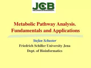
Receptors involving second messenger molecules. Many signals when bind to their receptors initiate a series of reactions in form of cascade reactions that at the end result in a specific intracellular response. The intracellular messenger systems function as signal amplification.
Receptors involving second messenger molecules • I. Adenylate cyclase system • Typical example is the adrenergic receptors β-receptors that bind to epinephrine and then trigger either an increase or decrease in the activity of the adenylate cyclase. • Adenylate cyclase convert the ATP into cAMP which act as second messenger and activate other enzymes to produce the activity. • Many signals (hormones) act through activating the cAMP like glucagon • the effect of signals on the second messenger is not direct but mediated through G-proteins. GTP-dependent regulatory proteins. The inactive form of G-protein binds to GDP while the active form bind to GTP. • The binding of hormone to its receptor activate the G-protein which affect the activity of adenylate cyclase • The activity of signal depends one the type of G-proteins. Gs stimulates the adenylate cyclase while Gi inhibits it. • The actions of G-protein _GTP complex are short lived because G-protein has an inherent GTPase activity, resulting in rapid hydrolysis of GTP to GDP and this causes the inactivation of G-protein.
Adenylate cyclase convert the ATP into cAMP which act as second messenger
The next link in the cAMP second messenger system is the activation of Protein Kinases by cAMP. • Family of enzymes called cAMP-dependent protein kinases. • Protein Kinase A is the typical example, consists of 4 monomers. 2 catlytics and 2 regulatory. • The active subunits catalyze the transfer of phosphate from ATP to specific serine or threonine residues of protein substrate. • the phosphorylated protein can act directly or can activate or inhibit other enzymes to produce the effect. • Not all protein kinases respond to cAMP, other types are cAMP-independent protein kinase like proetin kinase C. • The phosphate group can be removed by proetin phosphatases. • cAMP is rapidly hydrolyzed to 5-AMP by phosphodiesterase.
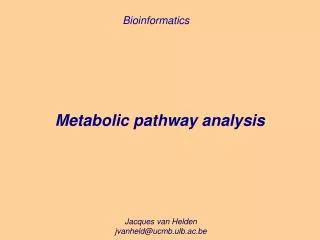
Role of Phosphatidylinositol 4,5 bisphosphate in Signal transduction
Ca+ is a second messenger in many signal transductions • Ca+ serves as a second messenger that triggers intracellular responses as exocytosis in neurons and endocrine cells, contraction in muscle and others • Ca+ is released from endoplasmic reticulum in response to signals (hormones, neurotransmitters) • Ca+ bind to Ca-binding proteins that called calmodulin • Calmodulin-ca complex binds and activates protein molecules usually enzymes, • Calmodulin is regulatory subunit of phosphorylase b kinase of muscle that activated by Ca activating the break down of glycogen. • Many enzymes are know to be modulated by Ca+ through calmodulin.
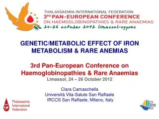
Nitric Oxide • NO act as endothelium relaxing factor, causes vasodilatation by relaxing vascular smooth muscle and also acts as neurotransmitters, prevents platelet aggregation and has a role in macrophage function. • It is highly toxic. Nitrous oxide ( NO2) the “ laughing gas” that used as anesthetic. • NO is very short lived and unstable converted into oxygen and nitrate and nitrite. • Synthesis of NO • NO synthase catalyzes the formation of NO from amino acid Arginine, FMN, FAD, and tetrahydrobiopterin are coenzymes for the enzymes • NO is synthesized in endothelial cells and diffuses to vascular smooth muscle activate the guanylate cyclase rise in cGMP which causes muscle relaxation. • Synthesis of NO is stimulated in the macrophages by bacterial liopolysaccharides activated macrophages form oxygen free radical that combine with NO to form compounds that are more bactericidal than NO itself
Other messenger systems • a. cGMP • b. Nitric oxide • * Cyclic guanosine monophosphate • It is analogous to cAMP pathway • Synthesized from GTP by guanylate cyclase, it an integral part of the receptor (not separated like Adenylate cyclase), it contains heme as prosthetic group and stimulated by nitric oxide. • Activate a spcific form of protein kinase called cGMP-dependent protein kinase also called protein kinase G • cGMP is hydrolyzed by phosphodiesterase • cGMP is a specialized messenger being involved in smooth muscle relaxation, platelet aggregation and the visual system. cAMP affects a wide variety of processes.
Nitric Oxide act as endothelium relaxing factor, causes vasodilatation by relaxing vascular smooth muscle and also acts as neurotransmitters, prevents platelet aggregation and many functions