Download

1 / 97
990 likes | 1.05k Views
Periodontal disease. Dentalelle tutoring. Question. Can everyone see and hear me OK? If you can’t – please text technical support at 519-859-4908. characteristics. Color

E N D
Periodontal disease Dentalelle tutoring
Question • Can everyone see and hear me OK? If you can’t – please text technical support at 519-859-4908
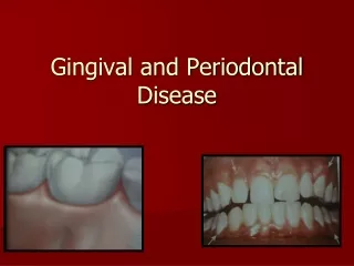
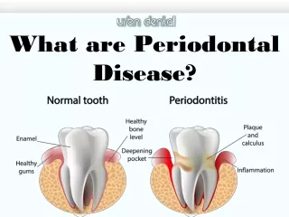
characteristics • Color Healthy gingiva usually has a color that has been described as "coral pink." Other colours like red, white, and blue can signify inflammation (gingivitis) or pathology. Although described as the colour coral pink, variation in colour is possible. This can be the result of factors such as: thickness and degree of keratinization of the epithelium, blood flow to the gingiva, natural pigmentation, disease and medications. Since the colour of the gingiva can vary, uniformity of colour is more important than the underlying color itself. Excess deposits of melanin can cause dark spots or patches on the gums (melanin gingival hyperpigmentation), especially at the base of the interdental papillae. Example of colors – red, pink, cyanotic • Contour Healthy gingiva has a smooth arcuate or scalloped appearance around each tooth. Healthy gingiva fills and fits each interdental space, unlike the swollen gingiva papilla seen in gingivitis or the empty interdental embrasure seen in periodontal disease. Healthy gums hold tight to each tooth in that the gingival surface narrows to "knife-edge" thin at the free gingival margin. On the other hand, inflamed gums have a "puffy" or "rolled" margin. Example of contours – bulbous, punched-out, flattened, cratered
continued • Texture Healthy gingiva has a firm texture that is resistant to movement, and the surface texture often exhibits surface stippling. Unhealthy gingiva, on the other hand, is often swollen and less firm. Healthy gingiva has an orange-peel like texture to it due to the stippling. Examples of texture – smooth, shiny, eroded, stippling • Consistency How the gingiva feels – healthy gingiva should be firm Examples of consistency – firm (fibrotic), spongy (edematous) • Reaction to disturbance Healthy gums usually have no reaction to normal disturbance such as brushing or periodontal probing. Unhealthy gums, conversely, will show bleeding on probing (BOP) and/or purulent exudate
Terms to be used • You need to determine the location, distribution and severity • Location – generalized or localized • Distribution– diffuse, marginal or papillary • Severity - Slight, moderate, severe
gingivits • Gingivitis is the mildest form of periodontal disease (gingivitis is NOT perio it CAN lead to perio if not treated).. It causes the gums to become red, swollen, and bleed easily. There is usually little or no discomfort at this stage. Gingivitis is often caused by inadequate oral hygiene. Gingivitis is reversible with professional treatment and good oral home care. • Factors that may contribute to gingivitis include, diabetes, smoking, aging, genetic predisposition, systemic diseases and conditions, stress, inadequate nutrition, puberty, hormonal fluctuations, pregnancy, substance abuse, HIV infection, and certain medication use.
Risk factors • AGE • Studies indicate that older people have the highest rates of periodontal disease. Data from the Centers for Disease Control and Prevention indicates that over 70% of Americans 65 and older have periodontitis. • SMOKING/TOBACCO USE • Tobacco use is linked with many serious illnesses such as cancer, lung disease and heart disease, as well as numerous other health problems. Tobacco users also are at increased risk for periodontal disease. Studies have shown that tobacco use may be one of the most significant risk factors in the development and progression of periodontal disease. • GENETICS • Research has indicated that some people may be genetically susceptible to gum disease. Despite aggressive oral care habits, these people may be more likely to develop periodontal disease. Identifying these people with a genetic test before they even show signs of the disease and getting them into early intervention treatment may help them keep their teeth for a lifetime. • STRESS • Stress is linked to many serious conditions such as hypertension, cancer, and numerous other health problems. Stress also is a risk factor for periodontal disease. Research demonstrates that stress can make it more difficult for the body to fight off infection, including periodontal diseases.
continued • MEDICATIONS • Some drugs, such as oral contraceptives, anti-depressants, and certain heart medicines, can affect your oral health. Just as you notify your pharmacist and other health care providers of all medicines you are taking and any changes in your overall health, you should also inform your dental care provider. • CLENCHING OR GRINDING YOUR TEETH • Clenching or grinding your teeth can put excess force on the supporting tissues of the teeth and could speed up the rate at which these periodontal tissues are destroyed. • OTHER SYSTEMIC DISEASES • Other systemic diseases that interfere with the body's inflammatory system may worsen the condition of the gums. These include cardiovascular disease, diabetes, and rheumatoid arthritis. • POOR NUTRITION AND OBESITY • A diet low in important nutrients can compromise the body's immune system and make it harder for the body to fight off infection. Because periodontal disease begins as an infection, poor nutrition can worsen the condition of your gums. In addition, research has shown that obesity may increase the risk of periodontal disease.
The Periodontium AB, alveolar bone; AC, alveolatrcrest; AM, alveolar mucosa; AP, alveolar process; CB, compact bone of alveolar bone proper; CEJ, cemento-enamel junction; CT, connective tissue; DEJ, dentino-enamel junction; ES,enamel space; G, gingiva; GE, gingival epithelium; GG, gingival groove; GM, gingival margin; GS, gingival sulcus; JE, junctional epithelium; MGJ, mucogingival junction; MS, marrow space; OE, oral epithelium; PDL, periodontal ligament; RCE, radicular (root) cementum; SE, sulcular epithelium;
The gingiva • Clinicians sometime use the terms "free" and "attached" gingiva. Although these terms may have some clinical relevance, they are anatomically incorrect. The determination as to whether the gingiva is "free" or "attached" is made by probing the gingival sulcus with a periodontal probe. • This instrument will frequently penetrate the junctional epithelium beyond the sulcus bottom, particularly in the presence of inflammation. This results in the clinical impression that the marginal gingiva is detached from the tooth to a much greater degree than is the case. Anatomically. "Attached" gingiva refers to the portion of the gingiva apical to the "free" gingiva which is firmly bound to the underlying tooth and alveolar process.
Fiber Groups These are largely composed of collagenous fibers. The dentogingival fibers (A) insert into the supracrestal root cementum and fan out into the adjacent connective tissue. Thedentoperiosteal fibers (B) insert into the supracrestal root cementum and blend with the periosteal covering of the adjacent alveolar process. The alveologingival fibers (C) insert into the alveolar crest and fan out into the adjacent gingival connective tissue. Thecircumferential fibers (D) follow a circular course around individual dental units. Thesemicircularfibers (E) insert on the approximal surfaces of a tooth and follow a semicircular course to insert on the opposite side of the same tooth. The transgingival fibers (F) insert into the approximal surface of a tooth and fan out toward the oral or vestibular surface. Theintergingival(G) fibers course along the oral or vestibular surfaces of the dental arch. Thetransseptalfibers (H) course from one approximal tooth surface to the approximal surface of the adjacent tooth.
Vessels and Nerves (i) Blood supply: The gingival blood supply originates from blood vessels in the periodontal ligament, the marrow spaces of the alveolar process and supraperiosteal blood vessels. These vessels in turn supply major capillary plexuses that are located in the connective tissue adjacent to the oral epithelium and the junctional epithelium. (ii) Lymphatics: The gingival tissues are supplied with lymphatic vessels that drain principally to submaxillary lymph nodes. (iii) Nerves: Branches of the trigeminal nerve provide sensory and proprioceptive functions. In addition, autonomic nerve endings are associated with the vasculature.
Supportive The periodontal ligament serves primarily a supportive function by attaching the tooth to the surrounding alveolar bone proper. This function is mediated primarily by the principal fibers of the periodontal ligament that form a strong fibrous union between the root cementum and the bone. The periodontal ligament also serves as a shock-absorber by mechanisms that provide resistance to light as well as heavy forces. Light forces are cushioned by intravascular fluid that is forced out of the blood vessels. Moderate forces are also absorbed by extravascular tissue fluid that is forced out of the periodontal ligament space into the adjacent marrow spaces. The heavier forces are taken up by the principal fibers.
Remodeling The periodontal ligament also serves a major remodeling function by providing cells that are able to form as well as resorb all the tissues that make up the attachment apparatus, i.e. bone, cementum and the periodontal ligament Undifferentiated ectomesenchymal cells, located around blood vessels, can differentiate into the specialized cells that form bone (osteoblasts), cementum (cementoblasts), and connective tissue fibers (fibroblasts). Bone- and tooth-resorbing cells (osteoclasts and odontoclasts) are generally multinucleated cells derived from blood-borne macrophages. *B = BUILDING, OSTEOBLASTS *C = TAKING AWAY BONE - OSTEOCLASTS
Sensory and Nutritive The periodontal ligament also serves a sensory function. The myelinated dental nerves that perforate the fundus of the alveoli rapidly lose their myelinated sheath as they branch to supply both the pulp and periodontal ligament. The periodontal ligament is richly supplied with nerve endings that are primarily receptors for pain and pressure. Finally, the periodontal ligament provides a nutritive function that maintains the vitality of its various cells. The ligament is well-vascularized, with the major blood supply originating from the dental arteries that enter the ligament through the fundus of the alveoli. Major anastomoses exist between blood vessels in the adjacent marrow spaces and the gingiva.
Cementum Cementum may be found both on the root as well as the crowns of teeth. It may also vary in its structure. Some forms of cementum may be cellular, while others are not. Some have a fibrillar collagenous matrix, while others do not. Cementum may be classified in the following ways: Radicular cementum: The cementum that is found on the root surface. Coronal cementum: The cementum that forms on the enamel covering the crown. Cellular cementum: Cementum containing cementocytes in lacunae within the cementum matrix. Acellular cementum: Cementum without any cells in its matrix. Fibrillar cementum: Cementum with a matrix that contains well-defined fibrils of type I collagen. Afibrillar cementum: Cementum that has a matrix devoid of detectable type I collagen fibrils. Instead, the matrix tends to have a fine, granular consistency.
Types 1. Acellular, afibrillar cementum This cementum is mostly composed of mineralized matrix, without detectable collagen fibrils or cementocytes. It is produced exclusively by cementoblasts. It is typically found as coronal cementum on human teeth. 2. Acellular, extrinsic fiber cementum This type of cementum has a matrix of well-defined, type I collagen fibrils. The fibrils are part of the, densely packed Sharpey's fibers, that are continuous with the principal fibers of the periodontal ligament. Because of their dense packing, the individual Sharpey's fibers that form the bulk of the matrix may no longer be identifiable as individual fibers within the cementum layer. This cementum, which is acellular, is located in the cervical two-thirds of the root of human teeth. It plays a major role in tooth anchorage.
3. Cellular, intrinsic fiber cementum This cementum contains cementocytes in a matrix composed almost exclusively of intrinsic fiber cementum. It is located almost exclusively at sites of cementum repair. It plays no part in tooth anchorage. However, it may be covered over by extrinsic or mixed fiber cementum, both of which are able to provide new anchorage. 4. Cellular, mixed fiber cementum It is found on the apical third of the root and in furcations (i.e. between roots). In these locations, the rate of cementum formation is usually more rapid than in the cervical region. The mineralized, extrinsic collagen fibers (Sharpey's fibers) run a more irregular course than in acellular, extrinsic fiber cementum. Intrinsic fibers are found interspersed among the extrinsic fibers of the cementum matrix, so that individual Sharpey’s fibers are more readily identifiable than in extrinsic fiber cementum. Cementoblasts are trapped in hollow chambers (or lacunae) where they become cementocytes. The thickness of radicular cementum increases with age. It is thicker apically than cervically. Thickness may range from 0.05 to 0.6 mm.
General The alveolar process is the portion of the jawbone that contains the teeth and the alveoli in which they are suspended. The alveolar process rests on basal bone. Proper development of the alveolar process is dependent on tooth eruption and its maintenance on tooth retention. When teeth fail to develop (e.g. anodontia), the alveolar process fails to form. When all teeth are extracted, most of the alveolar process becomes involuted, leaving basal bone as the major constituent of the jawbone. The remaining jawbone, therefore, is much reduced in height. The alveolar process is composed of an outer and inner cortical plate of compact bone that enclose the spongiosa, a compartment composed of spongy bone ( also called trabecular or cancellous bone). It is important to distinguish between the terms "alveolar process" and "alveolar bone" .
Alveolar Bone/Alveolar Process The alveolar bone proper lines the alveolus (or tooth housing) which is contained within the alveolar process. It is composed of a thin plate of cortical bone with numerous perforations ( or cribriform plate) that allow the passage of blood vessels between the bone marrow spaces and the periodontal ligament. The coronal rim of the alveolar bone forms the alveolar crest, which generally parallels the cemento-enamel junction at a distance of 1-2 mm apical to it
Fenestration Where roots are prominent and the overlying bone very thin, the bone may actually resorb locally, creating a window in the bone through which the root can be seen. This window-like defect in the bone is referred to as afenestration (F)
Dehiscence In some cases, as shown in this figure, the rim of bone between the fenestration and the alveolar crest may disappear altogether and produce a defect known as a dehiscence (D). Awareness of these defects is important when surgical flaps are reflected, as the exposure of such defects during surgery may aggravate their severity.
Bone Formation Bone is produced by osteoblasts (OB) that are found in the periosteum, endosteum and periodontal ligament adjacent to bone-forming surfaces. These specialized cells originate from less differentiated precursor cells close to the bone. These cells are in turn derived from undifferentiated ectomesenchymal cells found in the periosteum, endosteum and the periodontal ligament. During bone formation, osteoblasts become incorporated into bone as osteocytes (OC) that are completely surrounded by bone. The chamber in which they are trapped is called a lacuna (plur. lacunae). Osteocytes remain connected to osteoblasts and other osteocytes by cytoplasmic processes that run through small canals in the bone, or canaliculi (C).
Osteon Cortical plate of compact bone in the mandible. The mandible is enveloped by a well-developed cortex of compact bone. The bulk of the compact bone consists of cylindrical units of bone, the osteons or Haversian systems (HS). Each osteon has a central canal, the Haversian canal that houses a blood vessel. Haversian canals are linked to one another and the periphery of the cortex by Volkman canals that course perpendicularly to the Haversian canals. The outer and inner layers of the cortex consist of parallel lamellae of compact bone, called the external (ECL) and internal circumferential lamellae. The bone that fills the spaces between adjacent osteons is the interstitial bone.
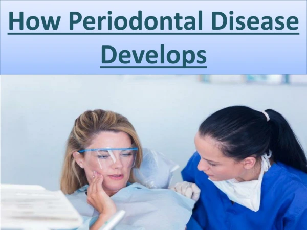
Periodontal disease • Untreated gingivitis can advance to periodontitis. With time, plaque can spread and grow below the gum line. Toxins produced by the bacteria in plaque irritate the gums. The toxins stimulate a chronic inflammatory response in which the body in essence turns on itself, and the tissues and bone that support the teeth are broken down and destroyed. • Gums separate from the teeth, forming pockets (spaces between the teeth and gums) that become infected. As the disease progresses, the pockets deepen and more gum tissue and bone are destroyed. Often, this destructive process has very mild symptoms. Eventually, teeth can become loose and may have to be removed.
A few forms: • Aggressive periodontitis occurs in patients who are otherwise clinically healthy. Common features include rapid attachment loss and bone destruction and familial aggregation. • Chronic periodontitis results in inflammation within the supporting tissues of the teeth, progressive attachment and bone loss. This is the most frequently occurring form of periodontitis and is characterized by pocket formation and/or recession of the gingiva. It is prevalent in adults, but can occur at any age. Progression of attachment loss usually occurs slowly, but periods of rapid progression can occur. • Periodontitis as a manifestation of systemic diseases often begins at a young age. Systemic conditions such as heart disease, respiratory disease, and diabetes are associated with this form of periodontitis. • Necrotizing periodontal disease is an infection characterized by necrosis of gingival tissues, periodontal ligament and alveolar bone. These lesions are most commonly observed in individuals with systemic conditions such as HIV infection, malnutrition and immunosuppression
Diabetes and periodontal disease • Diabetic patients are more likely to develop periodontal disease, which in turn can increase blood sugar and diabetic complications. • People with diabetes are more likely to have periodontal disease than people without diabetes, probably because people with diabetes are more susceptible to contracting infections. In fact, periodontal disease is often considered a complication of diabetes. Those people who don't have their diabetes under control are especially at risk. • Research has suggested that the relationship between diabetes and periodontal disease goes both ways - periodontal disease may make it more difficult for people who have diabetes to control their blood sugar. • Severe periodontal disease can increase blood sugar, contributing to increased periods of time when the body functions with a high blood sugar. This puts people with diabetes at increased risk for diabetic complications.
Gum disease and stroke • Several studies have shown that periodontal disease is associated with heart disease. While a cause-and-effect relationship has not yet been proven, research has indicated that periodontal disease increases the risk of heart disease. • Scientists believe that inflammation caused by periodontal disease may be responsible for the association. • Periodontal disease can also exacerbate existing heart conditions. Patients at risk for infective endocarditis may require antibiotics prior to dental procedures. Your periodontist and cardiologist will be able to determine if your heart condition requires use of antibiotics prior to dental procedures. • STROKE • Additional studies have pointed to a relationship between periodontal disease and stroke. In one study that looked at the causal relationship of oral infection as a risk factor for stroke, people diagnosed with acute cerebrovascular ischemia were found more likely to have an oral infection when compared to those in the control group.
women • PUBERTY • During puberty, an increased level of sex hormones, such as progesterone and possibly estrogen, causes increased blood circulation to the gums. This may cause an increase in the gum's sensitivity and lead to a greater reaction to any irritation, including food particles and plaque. During this time, the gums may become swollen, turn red and feel tender. • MENSTRUATION • Occasionally, some women experience menstruation gingivitis. Women with this condition may experience bleeding gums, bright red and swollen gums and sores on the inside of the cheek. Menstruation gingivitis typically occurs right before a woman's period and clears up once her period has started. • PREGNANCY • Some studies have suggested the possibility of an additional risk factor – periodontal disease. Pregnant women who have periodontal disease may be more likely to have a baby that is born too early and too small. However, more research is needed to confirm how periodontal disease may affect pregnancy outcomes. • All infections are cause for concern among pregnant women because they pose a risk to the health of the baby. The Academy recommends that women considering pregnancy have a periodontal evaluation. • MENOPAUSE AND POST-MENOPAUSE • Women who are menopausal or post-menopausal may experience changes in their mouths. They may notice discomfort in the mouth, including dry mouth, pain and burning sensations in the gum tissue and altered taste, especially salty, peppery or sour. • In addition, menopausal gingivostomatitis affects a small percentage of women. Gums that look dry or shiny, bleed easily and range from abnormally pale to deep red mark this condition. Most women find that estrogen supplements help to relieve these symptoms.
Chronic periodontal disease • Chronic periodontitis (a.k.a. adult periodontitis) is the most frequent type of periodontitis and is characterized by pocket formation and slowly progressing gum recession. • The condition may first appear in adolescence due to poor oral hygiene but in most cases it is after mid-30s when the clinical symptoms become significant. If chronic periodontitis symptoms are ignored, the loss of bone and gum tissue will lead to tooth loss. • It can be further divided into 3 stages; early, moderate and advanced stage.
Early periodontal disease • Periodontitis is the more dangerous form of periodontal disease. Infection and inflammation has spread to the bone supporting the teeth. When gum disease has progressed to the periodontitis stage, the supporting bone and fibers that hold the teeth in place start to get irreversibly damaged. The stages of periodontitis are defined as early, moderate, and advanced. The main factor for the classification to a certain stage of the disease is the degree of destruction of the supporting bone. • In the initial stage of periodontitis plaque bacteria continue to penetrate deeper between the teeth and gums. The environment becomes suitable for the establishment of anaerobic bacteria under the gums. Gingival pockets are formed below the gumline. • Greater inflammation and swelling of the gums • Gums begin to separate from teeth below the cemento-enamel junction • Gum bleeding when probing or brushing • Pocket depth up to 4mm • Infection reaches bone - Slight bone loss • Unpleasant breath or taste • Subgingival accumulation of plaque and calculus • Treatment of early periodontitis includes tooth scaling and root planing accompanied by improved oral hygiene. Despite the bone damage, the amount of bone loss in this stage of periodontal disease is minor so that usually no additional treatment is required.
Moderate periodontal disease • The surrounding connective tissues and alveolar bone become are seriously infected. Bacterial toxins and the body's enzymes fighting the infection break down the bone and connective tissue that hold teeth in place. First signs of tooth mobility appear due to bone loss. • Moderate Periodontitis • Gums recession – teeth appear longer • Root surface exposed - sensitivity - root decay • Persistent bad breath • Bleeding gums • Pocket depth of 5-6mm • Moderate bone loss (20 - 50%) • Periodontal abscesses may develop • Teeth may begin to loosen, drift and look separated • Moderate periodontitis is one of the most critical stages of periodontal disease, because some ‘damage control’ is yet possible before the condition reaches a phase when teeth can not be saved. Surgical treatments of gum disease can stop the progress of the disease but the damage is not reversible.
Advanced periodontitis • This final stage of periodontal disease is characterized by severe infection, loosening teeth and tooth loss. • Constant bad breath and bad taste • Spontaneous gums bleeding • Sensitive teeth due to exposed roots • Pocket depth over 7mm • Pus drainage in the mouth due to periodontal abscesses • Severe bone loss (more than 50%) • Teeth drifting out of place • Teeth become loose or fall out • Teeth become so mobile and the bone loss so severe that in many cases they can not be saved and have to be extracted. In other cases, teeth extraction is necessary in order to clear the infection. The advanced stage of periodontitis can be reached in some cases without intense visible alerting symptoms despite the severe underlying bone damage. • Extensive periodontal gum surgery that includes soft and hard tissue grafting are necessary in a treatment effort to save the affected teeth. Unfortunately the prognosis is not good if the condition has reached to this advanced stages of periodontal disease.
Aggressive periodontal disease • Aggressive periodontitis (a.k.a. early onset periodontitis) is a dangerous type of periodontal disease that can cause tooth loss in a short period of time. Common symptoms are the fast increase in the depth of periodontal pockets and the rapid loss of bone structure. • Sometimes the condition is localized affecting one or no more than 3 teeth in patients with good oral health. Generalized aggressive periodontitis affecting the whole mouth requires immediate treatment to prevent extensive tooth loss. Genetic factors and immune deficiencies are considered as causes of aggressive periodontitis in addition to the microbial etiology of gum disease. • Depending on the age when the condition first appears it used to be classified as: • Pre-puberty AP is a rare type of periodontal disease found in children less than 12 years old that can cause the loss of primary and/or permanent teeth. The disease is usually related to genetic disorders and may appear with the eruption of the first primary teeth. • Juvenile periodontitis begins at puberty and is defined by severe bone loss commonly limited around the first molars and incisors. It is more aggressive than other types of periodontal disease causing very rapid vertical bone loss across the teeth roots. Juvenile periodontitis is usually asymptomatic without the usual symptoms of gum disease such as gums inflammation or gum bleeding. • Rapidly progressive periodontitis occurs in the early 20s to mid-30s. Severe inflammation and rapid bone and connective tissue loss occur, and tooth loss is possible within a year of onset. • The goal of treatment in aggressive periodontitis is to fight the microbial etiology and (if possible) the contributing risk factors. Treatment methods are similar to those used for chronic periodontitis, including oral hygiene, tooth scaling and root planing, and periodontal gum surgery if needed.
Perio and systemic diseases • Periodontitis as a manifestation of systemic diseases is more often associated with younger age patients. Systemic conditions such as heart disease, leukemia, respiratory disease, and diabetes or disorders such as Down syndrome are associated with this form of periodontal disease. • A medical examination is required in cases of early-onset periodontitis to identify the presence of any systemic disease that might have triggered the periodontal disease.
anug • Acute Necrotizing Ulcerative Gingivitis (ANUG) is a severe and painful type of periodontal disease, which causes deep ulcerations of the gingival tissues. The condition is usually triggered by poor oral hygiene and poor nutrition. If left without treatment, the bacterial infection can lead to the necrosis of gum tissues and may spread to other areas of the body. • Acute Necrotizing Ulcerative Gingivitis is a much more severe condition than normal gingivitis, causing open gum sores and finally the death of the gum tissue.
continued • Pregnancy gingivitis is one of the most common dental problems during pregnancy. The condition is directly associated with the hormonal changes in the body of pregnant women. • Pericoronitisis a common dental problem of the gums in young adults at the age of 17-24 when the wisdom teeth normally erupt (break through the gum) in the mouth. It is a painful inflammation caused by the infection of the soft gingival tissues (gums) over or around a partially erupted tooth, most often a wisdom tooth. • Periodontal-endodontic lesions are another type of periodontitis that is related with infected hard tooth tissues except of the gums. In this condition an infection from a decayed tooth root may spread to the adjacent bone and gum tissues creating deep pockets around the tooth and leading to bone loss. • Desquamative gingivitis - one of the most painful but rare types of periodontal disease is a condition called desquamative gingivitis. The outer layers of the gums separate from the underlying tissue and peel away, exposing the inner layers and causing acute pain. This type of gum is more common in women after menopause.
Local antibiotic therapy • In this form of antimicrobial periodontal treatment the dentist places the antibiotic medication directly into the affected mouth tissues. The material dissolves slowly releasing a controlled dose of the antibiotic for a period of a few days. • There are several types of local antibiotic therapy, including: • Gel – A gel containing antibiotic is injected into periodontal pockets under the gums and sealed with a periodontal pack which is removed after 7 to 10 days along with any remaining gel. • Atridox is such a gel containing doxycycline. • Elyzol is a gel applied to the gum that is composed of metronidazole. • Powder — The dentist places the antibiotic in the form of a powder under the gums. The powder dissolves in about three weeks. • Chip - Chips containing antibiotic is placed under the gums and into the periodontal pockets. The chip dissolves over 7 to 10 days. • Periochip is a small piece of gelatin filled with chlorhexidine. • Actisite is a thread-like fiber similar to dental floss that contains tetracycline hydrochloride. • Arestin contains tiny round particles with the antibiotic minocycline.
Tetracycline Tetracycline administered as an adjunct to scaling and root planing has shown a pocket reduction greater than scaling and root planing alone. Mean pocket depth reductions were between 0.2 mm and 0.8 mm greater than scaling and root planing alone at six months (2.2 mm to 3.1 mm total reduction) in pockets 4 mm to 6 mm. Mean clinical attachment level gain at six months was 0.04 mm to 0.3 mm better than scaling alone (1.06 mm to 1.7 mm net gain). If surgical outcomes are included in the meta-analysis, then tetracycline with scaling and root planing with or without replaced flap surgery has a net attachment benefit over the intervention alone of 0.41 mm with a P = 0.003.
Types Periodontitis related abscess: When acute infections originate from a biofilm ( in the deepened periodontal pocket) Non-Periodontitis related abscess: When the acute infections originate from another local source. eg. Foreign body impaction, alteration in root integrity
Acute The abscess develops in a short period of time and lasts for a few days or a week. An acute abscess often presents as a sudden onset of pain on biting and a deep throbbing pain in a tooth in which the patient has been tending to clench. The gingiva becomes red, swollen and tender. In the early stages, there is no fluctuation or pus discharge, but as the disease progresses, the pus and discharge from the gingival crevice become evident. Associated lymph node enlargement maybe present.
Chronic This is the condition that lasts for a long time and often develops slowly. In the chronic stages, a nasty taste and spontaneous bleeding may accompany discomfort. The adjacent tooth is tender to bite on and is sometimes mobile. Pus may be present as also may be discharges from the gingival crevice or from a sinus in the mucosa overlying the affected root. Pain is usually of low intensity.
Rods Streptococcus viridans is the most common isolate in the exudate of periodontal abscesses when aerobic techniques are used. It has been reported that the microorganisms that colonize the periodontal abscesses are primarily Gram negative anaerobic rods. Althoughthey are not found in all cases of periodontal abscesses, high frequencies of Porphyromonas gingivalis, Prevotella intermedia, Fusobacteriumnucleatum, Campylobacter rectus, and Capnocytophagaspp have been reported.