Download
1 / 25
250 likes | 257 Views
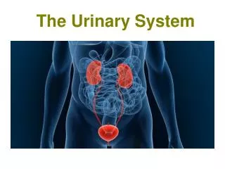
The Urinary System. Anatomy Ch. 15 The last chapter of notes for the year!. Function. The urinary system rids the body of nitrogenous wastes while regulating water, electrolyte, and acid-base balance of the blood. Kidneys. Filter fluid from the blood stream

E N D
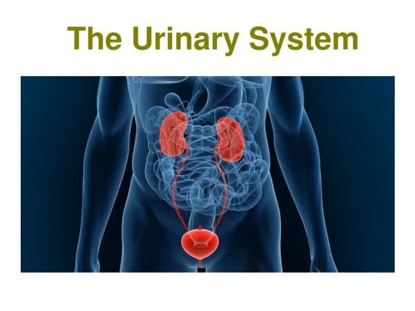
The Urinary System Anatomy Ch. 15 The last chapter of notes for the year!
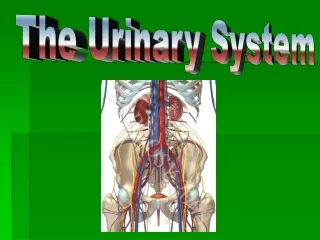
Function • The urinary system rids the body of nitrogenous wastes while regulating water, electrolyte, and acid-base balance of the blood.
Kidneys • Filter fluid from the blood stream • Dispose of nitrogenous wastes and excess ions • Regulate the blood’s volume and chemical makeup so that there is a proper balance between water and salts and acids and bases. • Produces the enzyme renin that regulates blood pressure • Produces the hormone erythropoietin that stimulates red blood cell production • Converts vitamin D to its active form called calcitriol which regulates levels of calcium in blood.
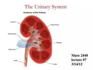
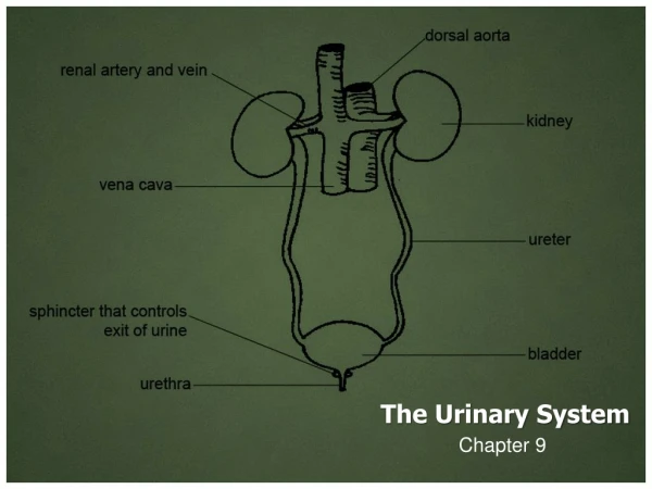
An adult kidney • 5 inches long • 2.5 inches wide • 1 inch thick • A region called the hilum is found on the medial side. The ureters, blood vessels, and nerves enter the kidney at the hilum. • The adrenal glands are found on top of the kidneys. They are functionally separate from the kidneys.
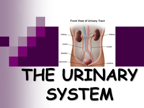
Major regions of the kidney • Renal Cortex: outer region • Renal Medulla: middle region • Renal pyramids: Triangle shape with a striped appearance found in the medulla. • The broad base faces the outside of the kidney and the apex faces the hilum. • The pyramids are separated by columns. • Renal Pelvis: inner region • Continual with the ureters • Extensions of the pelvis are called calyces and enclose the tips of the pyramids. The calyces collect urine.
Blood supply of the kidneys • The major artery that supplies blood to the kidneys are called the renal arteries (1). • As the renal artery approaches the hilum, it divides into segmental arteries (2). • Segmental arteries branch off into several interlobular arteries (3).These interlobular arteries travel through the renal columns until they reach the cortex. • Once these arteries reach the medulla-cortex boundary they become smaller arcuate arteries (4). These curve over the pyramids.
The arcuate arteries branch off into cortical radiate arteries (5). These arteries enter the cortex. • In the cortex, the blood that is in the arteries will move into afferent arterioles that enter a structure called the glomerulus. • The glomerulus is a dense ball of capillaries. The blood will leave the glomerulus through efferent arterioles. • After the blood leaves the efferent arterioles, it enters the peritubular capillaries. These capillaries surround tubes that collect substances the body needs. • The veins of the kidney trace the pathway of the arteries but in the opposite direction.
nephrons • Nephrons are the structural and functional units of the kidneys • There are over a million in each kidney • Responsible for forming urine • Each nephron contains 2 main structures: • glomerulus & renal tubule • There are 2 types of nephrons: • Cortical: located entirely in the cortex, majority • Juxtamedullary: located close to the cortex-medulla junction, loop of the nephron dips deep into the medulla
Glomerulus • The glomerulus is a bundle of capillaries • It is completely surrounded by a cup shaped structure called the Bowman’s capsule. • The Bowman’s capsule contains pores that allow for materials to pass through. • The Bowman’s capsule and glomerulus collectively make up the renal corpuscle.
Renal tubule • The first part of the tubule coils and is connected to the Bowman’s capsule. This region is called the proximal convoluted tubule (PCT). • The second part of the tubule that dips deeper into the cortex is called the loop of Henle. This loop forms a hairpin shape. • The last region of the tubule coils and is connected to a collecting duct. This region is called the distal convoluted tubule (DCT). • The DCT contains a region called the juxtaglomerular apparatus which secretes renin.
The coiling of the tubules allows for more surface area which increases reabsorption of necessary nutrients. • The PCT is called proximal because it is closest to the glomerulus. • The DCT is called distal because it is farther away from the glomerulus.
There are 2 capillary beds associated with each nephron. • The glomerulus is specialized for filtration (out). Due to high pressure, fluids and solutes are forced out of blood and into the Bowman’s capsule. • The peritubular capillaries are under low pressure and wrap around the entire tubule. These capillaries function in absorption (in).
Urine formation • Glomerular Filtration • The glomerulus acts as a filter. • Passive process. • Fluid passes from the blood into the Bowman’s capsule. • Once in the capsule the fluid is called filtrate and is essentially blood plasma without the blood cells or proteins. • Both proteins and blood cells are too large to pass through the filtration membrane.
Tubular Reabsorption • Besides wastes, filtrate contains many useful substances which must be reclaimed and returned to blood. • Tubule cells called transporters take up any needed substances from filtrate and pass them into extracellular space where they can be picked by up by peritubular capillaries. • The tubule cells are composed of simple cuboidal epithelium. • Some substances such as water pass through the membrane easily by osmosis. Other materials such as glucose and amino acids need carrier proteins which require energy. This is a type of active transport and requires ATP.
Nitrogenous wastes are poorly reabsorbed if at all. These substances tend to remain in the filtrate and are found in high concentrations in urine. • Examples • Urea: formed by the liver as an end product of protein breakdown • Uric acid: released when nucleic acids are metabolized • Creatinine: associated with creatine metabolism in muscle.
reabsorption • PCT • Location where most reabsorption occurs • water, glucose, amino acids, sodium, chloride, bicarbonate, phosphate, lactate, & citrate • Loop of Henle • Descending - water • Ascending – sodium, chloride, & potassium • DCT • sodium & chloride • Collecting Duct • water
Tubular Secretion • Tubular reabsorption in reverse • Some substances such as H+ and K+ ions and creatinine move from blood into the tubules to be released in urine. • This process is important for getting rid of substances such as certain drugs and excess ions, or as a means of controlling blood pH by removing H+ ions.
Urine formation • In a 24 hour period the kidneys filter between 150 and 180 liters of blood plasma. • During that same 24 hours, only about 1 to 2 liters of urine are produced. • Filtrate is not the same as urine • Filtrate contains everything blood plasma does except proteins and cells. • By the time filtrate reaches the collecting ducts, it has lost most of its water and almost all of its nutrients and ions. • Urine contains nitrogenous wastes and unneeded substances.
The normal light yellow color of urine is due to urochrome which is a pigment that results from the body’s destruction of hemoglobin. • The more solutes in the urine, the darker yellow color. • Dilute urine is a lighter color due to a higher concentration of water. • Urine is usually sterile and is slightly acidic with a pH of 6. • Solutes usually found in urine include Na and K ions, urea, uric acid, creatinine, ammonia, bicarbonate, and various ions.
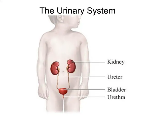
Ureters • The ureters are 2 slender tubes each 10 to 12 inches long and ¾ inches wide. • Each ureter runs from the renal hilum to the back of the bladder. • Each ureter is continual with the renal pelvis of the kidney at the upper end. • The ureters are passageways that carry urine from the kidneys to the bladder. • Smooth muscle in the walls of the ureters propel urine toward the bladder by peristalsis. • Once urine reaches the bladder it is prevented from flowing backwards by folds of the bladder mucosa.
Urinary Bladder • The urinary bladder is a smooth, collapsible, muscular sac the stores urine temporarily. • There are 3 openings to the bladder: 2 uretral openings in the back and 1 opening to the urethra. • The smooth triangular region of the bladder base outlined by these three openings is called the trigone. • Infections of the urinary tract tend to be located in the trigone area. • In males the prostate gland surrounds the neck of the bladder where it empties into the urethra.
The bladder wall contains 3 layers of smooth muscle. • The mucosa which is found on the inner layer of the bladder contains transitional epithelial tissue which allows it to change shape. • When the bladder is empty, it collapses to about 2 to 3 inches at the most and its walls are thick and folded. • As urine accumulates, the bladder expands to about 5 inches long and can hold between 500 and 1000 mL of urine.
Urethra • The urethra is a tube that carries urine by peristalsis from the bladder to the outside of the body. • There are 2 sphincter muscles found in the urethra. • The internal urethral sphincter is found at the bladder-urethra junction and is involuntary. • The external urethral sphincter found near the opening of the urethra and is under voluntary control. • In females the urethra is only about 1.5 inches long and only conducts urine. In males the urethra is about 8 inches long and conducts semen and urine.
Micturition • Micturition is the act of emptying the bladder. • The 2 sphincter muscles control the flow of urine. • Normally the bladder continues to collect urine until about 200mL have accumulated. • At this point stretch receptors in the wall of the bladder send messages to the sacral region of the spinal cord. • Messages are sent back to the bladder causing reflex contractions.
As the contractions become stronger, stored urine is forced past the internal sphincter into the upper part of the urethra. It is at this point when the person begins to feel like they need to go. • Because the external sphincter is under voluntary control, a person can choose to keep it closed and postpone urination. • Once the external sphincter is relaxed, urine is flushed from the body. • If a person continues to hold in the urine, the contractions will occur again when another 200 to 300 mL of urine accumulate. • Eventually micturition will occur whether the person wants it to or not.