Download
1 / 9
90 likes | 201 Views
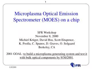
Detection of Perfluorinated Compounds By High-Pressure Microplasma Optical Emission Spectroscopy. SFR Workshop November 8, 2000 David Hsu, Michiel Krüger, Scott Eitapence, D. Graves, K. Poolla, C. Spanos, O. Solgaard Berkeley, CA.

E N D
Detection of Perfluorinated Compounds By High-Pressure Microplasma Optical Emission Spectroscopy SFR Workshop November 8, 2000 David Hsu, Michiel Krüger, Scott Eitapence,D. Graves, K. Poolla, C. Spanos, O. Solgaard Berkeley, CA 2001 Milestone: Build microplasma generating system. Test microplasma use as a sensor with bulk optical components.
Motivation • Microplasma features • Small, low power usage, simple to manufacture • High (atmospheric) pressure operation, intense excitation • Some applications may benefit from these unique features • Include microplasmas as part of an environmental sensor • Use microplasmas as a source for optical emission spectroscopy • Portable, inexpensive, versatile sensor • Test Case: Detection of C2F6 in argon at 700 Torr • PFCs are global warming gases, • Present in semiconductor mfg. exhaust at atmospheric pressure species, concentration ? OES
High-Pressure Microplasmas • Decreasing plasma length allows high-pressure operation of glow discharges • Microhollow Cathodes (MHC) Top View: Side View: 100s µm -V Metal Cathode Dielectric Metal Anode F • Microhollow Cathode Discharge (MHCD) forms in cathode hole • Circular geometry allows intense excitation and ionization in center of hole • pD ~ 10–20 Torr-cm
Experimental Setup • Microhollow Cathode • Electrodes, dielectric epoxied together • Hole mechanically drilled • Mo — Low work function, resistant to sputtering 200 µm Molybdenum Cathode Mica DielectricKAl2(AlSi3O10)(OH)2 500 µm Molybdenum Anode P 700 Torr 250 V, 8 mA -V 100 k Ocean Optics PC Card Spectrometer C2F6 0.15–3 sccm Ar 20 slm Pump (7–110 ppm C2F6 in Ar)
C2, C, F Transitions C II A3g–X’3 u F II F III C2 Swan v=0 C II F II F III F II C2 Swan v= -1 F II C2 Swan v=1 F II C II C2 Swan v=2 Spectral intensity increases noticeably with concentration. Broad molecular bands, sharp atomic peaks Integrate under peaks for intensity
Detection Limit from Molecular Bands C2F6 (20%) Intensity varies linearly with concentration Determine detection limit from where error bars hit x-axis
Detection Limit from Atomic Peaks C2F6 (±20%) Many lower intensity peaks, lower slopes than molecular bands
Total C, F, C2 Signal • Average over total C2, C, F signal to reduce errors • Lower error bars give detection limit of 4 ppm • Concentration error result of poor control of diluent flow 2
Conclusions • Detected 7 ppm C2F6 in argon at 700 Torr; Detection limit of 4 ppm C2F6 using total C, F, and C2 transition intensity • Suitable as atomic sensor; Identification of molecular species difficult • 2002 Milestone • Build micro-optics for spectral analysis. Complete preliminary designs for integrated MOES. • 2003 Milestone • Design and test integrated MOES. Calibration studies, sensor characterization. PFC (ppm) Diffraction grating