Download
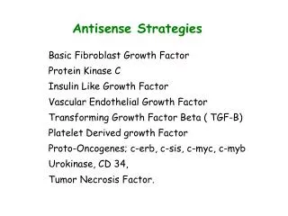
1 / 13
250 likes | 737 Views
Epidermal Growth Factor Receptor…. PDB ID: 1M14 (X-ray crystallography) . PDB ID: 1IVO (x-ray crystallography. Michelle McBride Molecular Anatomy Project . …is:. Picture here of structure diagram? Or next slide? . A transmembrane receptor glycoprotein

E N D
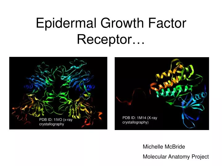
Epidermal Growth Factor Receptor… PDB ID: 1M14 (X-ray crystallography) PDB ID: 1IVO (x-raycrystallography Michelle McBride Molecular Anatomy Project
…is: Picture here of structure diagram? Or next slide? • A transmembrane receptor glycoprotein • A member of the EGFR or HER (Human Epidermal Receptor) Family • Associated with epithelial tissues… • …and therefore the following cancers: • Pancreatic -- Kidney • Ovarian -- Lung • Breast -- Prostate
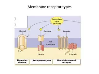
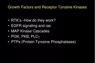
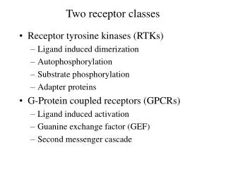
General Structure of EGFR • Extracellular domain • 4 domains: I and III bind to EGF, II is involved in dimerization and IV (often disordered) connects to trans membrane domain. • Trans membrane domain • A single alpha helix • Intracellular domain • Juxtamembrane domain • Tyrosine Kinase domain • N-lobe with the C-lobe form an ATP binding cleft. • C-lobe contains the Activation loop (A-loop), LVI motif and Catalytic site. • COOH terminal region thought to interact with kinase domain of neighboring EGFR family member to autophosphorolate.
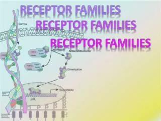
Step 1 EGF binding to extended (untethered) form of the extracellular domain Step 2 Dimerization of two bound EGFR extracellular domains Transfering the Signal into the Cell (Li, et al. 2005)
Extracellular Domain • Binding of EGF Bound extended form PDB ID: 1IVO (X-ray crystallography)
Extracellular Domain • EGF binds via 3 interacting sites.
Extracellular Domain The Beta hairpin arms of two bound EGFR molecules interacts by burying their hydrophobic surfaces in each other. • Dimerization PDB ID: 1IVO (x-ray crystallography)
Tyrosine Kinase Domain PDB ID: 1M14 (x-ray crystallography)
Tyrosine Kinase Domain • ATP Cleft
Cancer and EGFR’s Role in Apoptosis • Constitutive deregulation of EGFR is often observed in malignant tumors. • One mutation thought to cause such a deregulation is the truncation of the protein that lacks the extracellular domain. • Biological roles of EGFR • Progression of the cell cycle • Differentiation • In malignant cells, expression of EGFR leads loss of the ability of the cells to polarize and therefore differentiate. • Cell survival. • In cells that have been detached from cellular matrix, EGFR provides a measure of protection from apoptosis. • Cell migration • In tumors, cells are able to survive (resist apoptosis) without intracellular or matrix attachments, allowing them to survive migration.
Intracellular matrix and other cell attachments work with growth factor receptors like EGFR to resist apoptosis when stressed. EGFR and Apoptosis Tumor cells that have lost intracellular matrix connections as well as cell/cell connections depend solely on growth factor receptor cell survival signals Whether blocked or unblocked cells with cell/cell adhesion are able to survive.
What happens when… ….stressed tumor cells are EGFR blocked? EGFR MAPK Bcl-xL (anti-apoptotic protein) (other members of Bcl family that promote apoptosis are unaffected by this mechanism) STAT3 also becomes activated by EGFR contributing to increased Bcl-xL expression. In normal cells EGFR does not phosphorolate STAT3.
References • 1.Stamos, J., Sliwkowski, M.X., Eigenbrot, C. (2002) Structure of the epidermal growth factor receptor kinase domain alone and in complex with a 4-anilinoquinazoline inhibitor. J.Biol.Chem. 277: 46265-46272. • 2. Choowongkomon, K., Carlin, C.R. (2005) A Structural Model for the Membrane-bound Form of the Juxtamembrane Domain of the Epidermal Growth Factor Receptor J.Biol.Chem. 280: 24043-24052 • 3. Ogiso, H., Ishitani, R., Nureki, O., Fukai, S., Yamanaka, M., Kim, J.H., Saito, K., Inoue, M., Shirouzu, M., Yokoyama, S. (2002) Crystal Structure of the Complex of Human Epidermal Growth Factor and Receptor Extracellular Domains. Cell (Cambridge,Mass.) 110: 775-787 • 4. Li, S., Schmitz, K.R., Jeffrey, P.D., Wiltzius, J.J.W., Kussie, P., Ferguson, K.M. (2005) Structural basis for inhibition of the epidermal growth factor receptor by cetuximab Cancer Cell 7: 301-311