Download
1 / 59
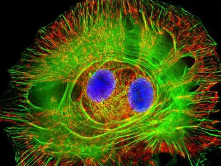
590 likes | 708 Views
Letting someone else doing my job. http://probes.invitrogen.com/resources/education/tutorials/1Intro/player.html. Epi-illumination is form of Kohler Illumination: Objective is also condenser. White light (regular Kohler) Brightfield, phase, etc. Light is focused At back aperture

E N D
Letting someone else doing my job http://probes.invitrogen.com/resources/education/tutorials/1Intro/player.html
Epi-illumination is form of Kohler Illumination: Objective is also condenser White light (regular Kohler) Brightfield, phase, etc Light is focused At back aperture Of the objective, Conjugate to condenser aperture Different illumination And image paths Lamp or laser lens detector Detect at 90 degrees Split with dichroic mirror Greatly increases S/N
Epi-illumination separates light source, Fluorescence signal Second barrier filter Selects signal From background First barrier filter Selects excitation Arc lamp dichroic mirror objective lens specimen
General Jablonski Diagram Typical molecular timescales: Absorption: instantaneous (10-17 s) Vibronic Relaxation ~ 10-12 s Fluorescence: 10-6 -10-12 s 10-9 most typical Singlet to a singlet (strong) Phosphorescence: 10-3 -10-6 s Triplet to singlet (weak) S2 T1 S1 S0 Fluorescence always from relaxed level of S1
Magnitude of Extinction Coefficients Extinction coefficient ε: Beer’s Law A= εcl Strong absorbers (dyes) have ε between 20,000-100,000 Absorption cross section is also used: 1x 10-16 cm2 = 23,000 ε Brightness= absorption coefficient* QY Oscillator strength is integral of the absorption band Sum rule: oscillator strength, f, for one electron over all transitions is: 1x 10-16 cm2 eV
Emission spectrum is independent of of path excitation (both spectral wavelength and width): always from S1 Emission intensities will be different Due to different absorption probability: Franck-Condon Principle
Franck-Condon Principle Consider electronic states anharmonic oscillators: bond length Most probable transitions are “vertical” Big geometry change=broad spectrum (smaller maximum absorption) Both for absorption and emission: conserve oscillator strength
Geometry and Absorption and Emission Spectra Bigger Stokes shifts provide better signal to noise In fluorescence because of filter efficiencies, (dichroics, low pass, high pass) But usually involve large geometry change: lower intensities Spreading out oscillator strength
Fluorescence Quantum Yieldφ: important for dyes Ratio of emitted to absorbed photons Quantum Yield: (k is rate, Inverse of time) Measured lifetime is sum of natural lifetime and non radiative decay paths Lifetime is useful contrast Probe of environment
B. Fluorescent Probes • Molecular Probes (Invitrogen) • www.probes.com • Catalog contains thousands of fluorescent probes, with valuable technical information.
Immunofluorescence Imaging – Detect Proteins IgG Fab Fc
Fluorescence in situ Hybridization (FISH) – Detecting Nucleic Acids
Fluorescein: most common dye for microscopy Bluegreen Xanthene family “green fluorophore” e=80,000, φ~0.9 • High quantum yield • General purpose • But degrades quickly • Small Stokes shift • (filter bleedthrough) Brightness ~eff Many functionalized forms for cell imaging: pH, ion sensing
Rhodamine 6G GreenRed Xanthene family “red fluorophore” • High quantum yield • General purpose • Good stability • Also Small Stokes shift Internal Donor-acceptor pair Red-shifts the spectra relative to fluorescein Many functionalized forms for cell imaging
Green Fluorescent Protein (GFP) Fluorophore made of Ser65, Tyr66 and Gly67 • Requires no co-factor or substrate. • Works in almost any organism. • Easy to quantify. • Genetically modifiable. Tsien, Ann.Rev. Biochem.67, 509 (1998)
Many fluorescent proteins: Jellyfish, Coral Reefs
Colored Proteins allow labeling of multiple specific organelles
Variants of Fluorescent Proteins BFP EBFP, Sapphire, T-sapphire CFP ECFP, mCFP, Cerulean, CyPet, AmCyan, Midoriishi Cyan GFP EGFP, Azami Green, TurboGFP, ZsGreen, Emerald YFP EYFP, Topaz, Venus, mCitrine, YPet, ZsYellow1, PhiYFP OFP mBanana, Kusabira Orange, mOrange RFP dsRed, tangerine, dTamato, mStrawberry, AsRed2, mRFP, mCherry, mRasberry, mPlum, JRed, HcRed
GFP Chromophore Aequorea: FSYGVQ Renilla: FSYGDR p-hydroxybenzylidene-imidazolidone Ser - dehydroTyr - Gly
Chromophore Maturation Takes ~ 30 min for wild type GFP.
GFP cellular protein cellular protein GFP GREEN FLUORESCENCE PROTEIN Jelly fish isolate DNA encoding GFP couple gene for GFP with gene for protein of interest transform cell with altered protein Completely general and versatile
Problems with Fluorescent Protein • Size comparable to the target. Might interfere with the function of the target protein • Maturation time • Probably not 100% fluorescent • PH dependence • Many variants mis-fold when fused to another protein
Linearly Polarized Light s= horizontal p= vertical For propagation Parallel to floor
Polarizer is device that selects polarization Can be crystal or film (Polaroid) Operation of Analyzer (Birefringent) Light transmitted at angle relative to angle of 2 Crossed polarizers
Combining linear polarized light OUT OF PHASE IN PHASE Elliptical Polarization Linear Polarization
Circularly Polarized Light • Decompose to linear polarized light with 1/4 phase shift. • No direction (always pass 50% through polarizer in dependent of polarizer orientation) • NOT the same as unpolarized light. • Can be converted back to linear polarized light with birefringent materials (1/4 wave plate).
Absorption is polarized Fluorescence is also polarized GFP Crystal
Anisotropic sample - Fluorescent intensity is dependent on the polarization _and_ the orientation of the molecules Isotropic sample • Fluorescent intensity is independent of excitation polarization • Fluorescence is polarized if the excitation is polarized. Fluorescence anisotropy r < 0.4
Microscopic Measurements of Anisotropy r = r0 / ( 1 + / ) Use Small Numerical Aperture
Fluorescence Resonance Energy Transfer (FRET) Förster Radius The distance at which energy transfer is 50% efficient (i.e. 50% of excited donors are deactivated by FRET) is defined by the Förster Radius (R0).
Survey of FRET-Based Assays • Protease activity • Calcium Ion measurements • cAMP • Protein tyrosine kinase activity • Phospholipase C activity • Protein kinase C activity • Membrane potential
FRET probes conformational changes Different conformation gives Different FRET signature
Inter and Intramolecular Forms of FRET with Proteins CFP-YFP good combo FRET increases In both cases Protein-Protein Interactions In cytoplasm and membranes
When FRET Occurs No FRET for No overlap of donor emission, acceptor absorption No FRET for Orthogonal dipole orientation No FRET for molecules more than 10 nm apart
Fluorescence Resonance Energy Transfer - Detection of Probe Proximity R0 typically 40-50 Angstroms 50% transfer
Donor Acceptor Ro (Å) Fluorescein Tetramethylrhodamine 55 IAEDANS Fluorescein 46 EDANS DABCYL 33 Fluorescein Fluorescein 44 BODIPY FL BODIPY FL 57 Fluorescein QSY 7 dye 61 Cy3 Cy5 53 CFP YFP 50 Typical Values of Ro green red GFPs and other colored “FPs have transformed FRET microscopy
FRET Considerations: 1. Spectral overlap 2. Chromophore orientations 3. Distance dependence (Eff. 1/R6) 4. How to quantify?
Practical Challenges to FRET Quantitation • Emission from A contaminates D channel (filters) • Emission from D contaminates A channel • Unknown labeling levels for D and A • Signal variation due to bleaching • Complicates kinetic studies • Bleaching rate of D can actually be slowed by FRET • Solutions: • Separately labeled D and A controls to define bleedthrough • Acceptor destruction by photobleaching to establish • Dual wavelength ratio imaging to normalize away variations in label levels and bleaching effects
Ca2+ Release During Shrimp Egg Activation • From Lindsay et al. (1992). Extracellular Mg2+ Induces an Intracellular Ca2+ Wave During Oocyte Activation in the Marine Shrimp Sicyonia ingentis. Dev. Biol. 152:94-102.
Low quantum yield with no Ca2+, big increase When binds Ca2+:up to 50 fold increase Not absolute concentration of ions, measure relative changes: easier Fluo- dyes By Tsien Choose depending on desired Range of sensing
Blue Ca2+ Indicators: Fluo-3 has single Ex and Em wavelengths • A visible light excitable dye (488 nm), so Argon laser can be used. • Emission at 525 nm. • OK for qualitative detection but not quantitative.
Indo-1 Calcium Sensing Ratiometric using single excitation, dual emission Excite 338 nm, collect 405, 485 nm fluorescence Determine absolute calcium concentration by imaging